High time for a check up

by Bethany Palumbo, life collections conservator

This month marks three years since the completion of our ‘Once in a Whale’ project. The initial conservation undertaken in 2013 focused on the cleaning and stabilisation of five whale skeletons, which had hung from the roof of the Museum for over 100 years.

The skeletons were lowered into a special conservation space, where the team were able to work up close with the specimens. As well as the cleaning, they improved incorrect skeletal anatomy, replacing old corroded wiring with new stainless steel. For final display, the specimens were put into size order and rigged using new steel wiring, with the larger specimens being lifted higher into the roof space to make them a more prominent display than previously. You can read all about the project on our blog, Once in a Whale.

Three years on, our conservation team felt it was a good time to check on the specimens to see how they’re coping, post-treatment, in the fluctuating museum environment.

Conservation intern Stefani Cavazos works on high to clean the Beaked Whale

It’s been wonderful to see the whales on display and their new position looks very impressive. However, when the time came for making this recent conservation assessment, the new height was greater than any of our ladders could reach. Specialist scaffolding was brought in to allow the conservators to access the specimens. Starting at the highest level, with our Beaked Whale, cleaning was completed using a vacuum and soft brush for delicate areas. This removed a thick layer of dust and particulate debris: especially satisfying work!

Dust gathered on the Beaked Whale fin

With cleaning complete, visual assessments could then be undertaken. These showed that while the specimens were still very stable, a few areas of bone have continued to deteriorate, visible in cracking and flaking of the surface. In other areas, the fatty secretions which we previously removed using ammonia had once again started to emerge. We had expected to see this though, because, in life these whales’ bodies contained a lot of fat, deep within the bones and this is notoriously impossible to completely remove.

Lubricant stain seen on a vertebra

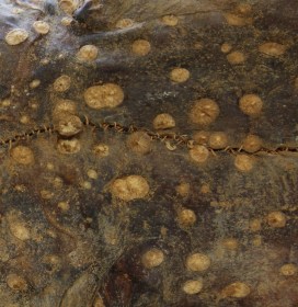
It was also observed that the lubrication used on the new rigging bolts had melted and dripped down the wires. You can see in the photo above how this has become drawn into the vertebrae of the Orca and Common Dolphin, staining them yellow. While no conservation treatment was undertaken due to time restrictions, thorough photography was performed to document these changes and once time permits this can be carried out.

This shows how conservation work, especially with natural history specimens, is a gradual, ongoing process. With frequent check-ups and specialist attention, these whales will be able to continue their life as our beautiful display specimens.

Stories from the Stores

by Hannah Allum, Move Project Manager

Museums are space-hungry places. There’s rarely enough room to properly house the collections we currently hold, never mind finding space for new material. Roughly 90% of museum collections are not on public display, so many museums use storage beyond their main building, in offsite stores.

The Museum of Natural History has faced the same challenges and, for the past few decades, has found a solution in a somewhat surprising building. Where might you least expect to find a vast collection of rocks and fossils dating back millions of years? How about a nineteenth century church, complete with beautiful stained glass windows?!

Stained glass windows in the former church

As atmospheric as it is, this deconsecrated church is no place for good quality museum storage. To do the collections justice they need a monitored and controlled environment, suitable containers and pest prevention measures. Plus, it’s always a bonus to be able to access specimens without playing a very nerve-wracking game of Jenga or Tetris!

Carving detail in the former church

So, once again we are faced with the most terrifying and exciting of museum tasks; a collections move project! In 2016 we successfully relocated approximately 17,000 specimens from some of our old offsite stores and showed you some behind the scenes secrets along the way with the hashtag #storiesfromthestores. This time we have a team of eager beavers (also known as project assistants) who can’t wait to share glimpses of the project as we go, so make sure to keep an eye on @morethanadodo on Twitter.

An oats box made an unusual storage solution for Earth materials

After a much needed clean of the church, we’re starting to prepare the material to be moved. Alongside the specimens, we’re already coming across some interesting historical packaging. It seems that the mass consumption of Oxo cubes was a pre-requisite for collectors of old, as many specimens are stored in these retro tins. A few other repurposed boxes include; Quaker oats, Sharp’s toffee, Fry’s chocolates, marmalade, mustard oil and even wine crates. Quite the picnic!

A toffee tin for storing fossils – just one of the interesting choices for collections tins over the years.

Once removed from the church, the project team will be working on processing these collections in a dedicated space, as well as giving them some TLC and new homes (boxes). This will take place at a specialist heritage storage site until the end of 2018. Keep your eyes peeled for more on the treasures from this trove.

Paint it green

In the process of researching or conserving old pinned insects, it’s common to find a green deposit clustered around the pin. This is known as verdigris and is a natural patina created when the metal oxidizes over time. Katherine Child is Image Technician in the Museum’s Life collections and takes photos of insects for researchers, students, artists and publications. She is also an artist in her own right, so when she witnessed verdigris being removed during a conservation project, she came up with an inspired idea.

A clearwing moth before conservation, showing verdigris spreading where the metal and the insect fats, or lipids, react.

A few years ago I read a book called Colour: Travels Through the Paintbox, by Victoria Finlay, and was interested to learn that verdigris was once used as a pigment. Verdigris, which I now know translates from French as ‘Green of Greece’, is a word that’s been in my vocabulary since I was small.  I loved its rich bright blue-green colour, which is often seen on old copper piping or copper statues.

Verdigris forms when copper or a copper alloy reacts with water, oxygen, carbon dioxide or sulphur.

L: Three years’ worth of verdigris, ground and ready to make into paint.
R: A second attempt at mixing the paint, this time using linseed oil.

As early as 5thcentury AD, it was used in paint-making, and until the late 19th century it was the most vibrant green pigment available. But it was unstable – Leonardo da Vinci warned that it ‘vanishes into thin air if not varnished quickly.’ These days synthetic pigments provide a more constant alternative.

Despite its past uses, verdigris is a big problem in pinned insect collections. Nowadays stainless steel pins are used, but pins containing copper still remain in old collections and these react with air and insect fats. The more fatty the insect, the more verdigris tends to form and, if left, it can damage a specimen irreparably.

Comprising around five million or so insects, the Hope Entomological Collections here in the Museum take quite a bit of looking after. A few years ago a project to catalogue and conserve many of its butterfly and moth specimens was undertaken and the removal of verdigris and repining of insects was part of this.

With paint-making in mind, I asked that the beautiful, but problematic, substance be saved.  About three years on I finally got around to using the pigment, which I had also been adding to while photographing the collections.

I chose a variety of differently shaped moths to paint (most of the verdigris came from moths, so moths seemed the most apt subject). To narrow my options further I went for green moths. Some of the specimens I chose had verdigris on their pin, so I was able to take pigment and use it to paint the very specimens from which it came!

Katherine tested out the newly made verdigris paint in her sketchbook.

After a first failed attempt to make watercolour paint (during which pigment and water remained stubbornly separate due to the greasy insect fats still present), I tried again, this time using linseed oil to make oil paint – and it worked! Traditionally a flat bottomed tool called a muller was used to press pigment into the water or oil. Not having one of these, I used the flat end of a pestle and a mortar which did the trick.

A Miscellany of Moths, the finished verdigris painting.

The paint went surprisingly far and, following on from the 14 green moths, I plan to use up the remainder to paint beetles.

Katherine’s Miscellany of Moths painting can be seen on display in the Museum’s Community Case until 18th October.

A rare beetles turns 21

by Darren Mann, Head of Life Collections

Many years ago, when re-identifying dung beetles in the collections of the British Entomological and Natural History Society, I found a specimen that I didn’t immediately recognise. So I borrowed it, and after a few hours of checking the European literature back in Oxford, I realised that I’d found a beetle that had not been recorded anywhere in Britain before.

The small black circles show the locations of known records for Melinopterus punctatosulcatus.

The dung beetle in question was Melinopterus punctatosulcatus, a species widely distributed across Europe but until this discovery unknown in Britain, despite its presence in the BENHS collection. This is because it had been misidentified as a different species: the beetle superficially looks like two closely-related species, and so had been overlooked by beetle collectors for over a hundred years.

Since that initial specimen, I have scoured numerous UK museum collections and to date have found a total of just 20 specimens, distributed across the World Museum in Liverpool, the National Museum Wales in Cardiff, and here in the Museum of Natural History in Oxford. All these specimens are from Deal, Kent and were caught between 1891 and 1910.

The last known record is of a single specimen from Ryarsh, Kent collected in 1938, which just happens to be the first specimen I found some 20 years ago in the BENHS collection.

The male genitalia of Melinopterus punctatosulcatus. The appearance of the genitalia is one of the best ways of identifying one species of beetle from another.

But this week, the 21st known specimen was discovered in our collections by Mary-Emma, a placement student who is with us from the University of Reading. She uncovered the beetle during the re-curation and identification of a collection made by A. J. Chitty. Thankfully the specimen was a male, so we were able to confirm the identification using the genitalia – one of the best ways of determining a species.

It seems that Mr Chitty had a knack for finding this particular species of dung beetle, since 14 of all the known specimens were caught by him at Deal. It’s just a shame that he didn’t realise his amazing discovery at the time.

Mary-Emma identifies Melinopterus punctatosulcatus by examining the dissected genitalia, visible on the right hand side of the monitor screen.

In the recent Conservation Status Review of dung beetles, Melinopterus punctatosulcatus was designated as Regionally Extinct in the UK because there have been no known sightings since that one in 1938. So this species possibly went extinct in Britain before we even realised that it was here. And were it not for museum collections we may never have known it once lived in Britain at all.

Solving a prickly mystery…

oumnh-zc-17347_echinorhinus_brucus_mark_carnall_01-800px

by Mark Carnall, Life Collections manager

Visitors to the Museum often comment on just how many things there are to see here, but in fact only a small percentage of the collection is on public display – less than 0.1%. The remaining 99.9% is held in storage for use in research, teaching and to loan to other museums and universities.

I receive hundreds of enquiries from researchers all over the world who are interested in many aspects of animal biology. In exchange for our information about the skeletons, skins, specimens preserved in fluid, nests, shells and taxidermy here, the Museum gets knowledge from world experts who are publishing new science.

Bramble Shark (Echinorhinus brucus)
Bramble Shark (Echinorhinus brucus). Image: Illustrations of South Africa / Wikimedia

One such recent enquiry, from researchers Dr Samuel Iglésias and Frederik Mollen, concerned the Bramble or Spinous Shark (Echinorhinus brucus). You may not have heard of the Bramble Shark – it’s an enigmatic bottom-of-the-sea dwelling species – but this is one of the reasons why the researchers are trying to track down all known specimens held in European institutions.

The characteristic brambles of the Spinous Shark, Echinorhinus brucus

Bramble Sharks are so called because of the large thorn-like structures, called dermal denticles, that cover the skin. These sharks are thought to be once common in European waters but are now virtually extinct.

I checked the Museum databases and stores and found 14 Bramble Shark specimens in the collections: one taxidermy specimen; one set of jaws with skin; a section of skin; and 11 fluid-preserved dissections. Unfortunately, as is often the case in older natural history collections, the fluid specimens did not have data associated with them – information such as age, locality, who collected them, what the dissections were or who dissected them.

After passing images and this list of specimens to the researchers they sent through an 1875 paper titled The Brain and Cranial Nerves of Echinorhinosus spinosus with notes on the other viscera by Bruce Clarke and the excellently named Hatchett Jackson. This paper describes the dissection of two specimens at the University of Oxford, one of which was a female collected from Penzance on 15 February 1875.

Armed with this new information I was able to cross-reference this dissection with some of our archives to confirm that most of the dissected specimens actually came from this single female shark collected in 1875, which was dissected and the parts used in illustrations in a technical publication.

Section of skin previously identified as Bramble Shark but now re-identified as from a Porcupine Ray (Urogymnus asperrimus)

Another discovery came while photographing the skin specimen, and comparing it to the other dry specimens. It became clear that the thorns of the skin were very different to the typical Bramble Shark arrangement and it turned out that the skin was in fact from a Porcupine Ray (Urogymnus asperrimus).

The happy ending here is that the researchers now have better information for their publication and these specimens will be put ‘on the map’ in the technical literature for other researchers to access.

Although the Museum collections are effectively one Bramble Shark down (but up one Porcupine Ray skin), we do now have better information about the age, locality, relationship, identification, citation in the literature, and history of some these specimens. This makes them even more useful for research in the future.

Not all enquiries end with such a deeply satisfying result, but it is certainly nice when they do.

Darwin, dolphins and a ‘Monkeyana’

macmanimalkingdompl60-crop-750

The Museum is home to a vast collection of natural history specimens but is perhaps less well-known for its substantial art and object collection. This material became the focus of Charlie Baker and Imogen Stead, two of our summer interns, as they spent six weeks researching, organising and curating it for the Museum.

The range and amount of material was formidable: numerous prints, non-scientific objects, paintings, photographs and sculptures from across the Museum, all coming together into a single organised collection for the first time. Here, Charlie and Imo unearth just a small sample of some of the items they catalogued during their time at the Museum:

Nautilus Imperialis
This beautiful print shows a fossil of the Nautilus Imperialis. It is one of the largest prints the Museum holds: measuring 48cm x 43 cm, it’s too large for the scanner! It has a small pamphlet of text stuck to it, just visible in the picture, and we speculate that this may have been promotional material for James Sowerby’s Mineral Conchology of Great Britain, the first volume of which was published in 1812, the same year as this print.

nautilusimperialis-for-web
Nautilus Imperialis print

Plate 60 from The Animal Kingdom
This is a plate from Henry MacMurtrie’s translation of Georges Cuvier’s Le Règne Animal, showing a few species of the genus Delphinus, or Common Dolphins. The Museums has 32 plates from this book in the collection. The publication demonstrates the intellectual collaboration between countries and the international appeal of Cuvier’s famous work. In fact, the collections holds plates from the original French and two different English translations.

macmanimalkingdompl60
Plate 60 from ‘Animal Kingdom‘ (Le Régne Animal) by Georges Cuvier, 1829

Slide cutter
This tool was found in a chest of drawers in the Hope Library at the Museum. It initially baffled us, but staff identified it as a slide cutting tool. The circular blade scores the glass, and the notches are used to carefully break off the piece of glass. When cataloguing and storing it, we discovered the blade is still sharp enough to cut through a sheet of paper!

slidecuttercloseup
Slide cutter tool

Photo of Charles Darwin
This framed photo of the great Victorian scientist is one of 22 pieces of art hanging in the Museum’s Hope Library. What makes this copy of the photo special, however, is the caption beneath it: “I like this photograph very much better than any other which has been taken of me.”

darwin
Photograph of Charles Darwin with his annotations

Monkeyana Cartoon
One of the most bizarre items we came across was this 1828 satirical cartoon about lawyers. The collection has eight ‘Monkeyana’ cartoons, all by Thomas Landseer.

monkeyana
Monkeyana Cartoon by Thomas Landseer, 1828