When life got hard

By Dr Duncan Murdock, Research Fellow

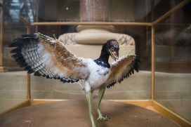
Whether you’re a great white shark with a deadly conveyor belt of teeth, a deep sea snail with a coat of armour or a coral building the Great Barrier Reef one polyp at a time, mineralized skeletons are a crucial part of many animals’ way of life. These hard skeletons – shells, teeth, spines, plates and bones – are all around us.

The fossil record is full of the remains of the skeletons of long-extinct critters, so much so that entire layers of rocks can be composed almost completely of them. But this has not always been the case…

A piece of 425 million year old sea floor containing the skeletons of trilobites, brachiopods, bryozons, corals and gastropods preserved as limestone

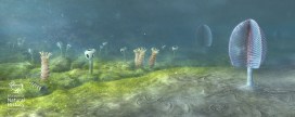
Travel back some 570 million years to a time known as the Ediacaran and the picture is very different. Although there were large-bodied creatures that were possibly animals, they were entirely soft-bodied. Then, right at the end of the Ediacaran Period, the first animals with hard skeletons evolved, creating strange tubes, stacked cones, and other bizarre forms such as Namacalathus, which resembles a baby’s rattle!

Some of the first animals with skeletons, Cloudina and Namacalathus alongside the soft-bodied Ediacaran fauna. Reconstruction based on rocks from Namibia, Southwest Africa, from 543 million years ago. Image: Mighty Fossils.


In the following few tens of millions of years, in the early part of the Cambrian Period, a whole host of animals burst onto the scene baring their ‘teeth’, hiding in their shells, and bristling their spines. In fact, we can trace the origin of almost every kind of animal skeleton to this relatively short window of the Earth’s past.

In my research, I have compiled the evidence for how and when these skeletons first appear. Three key observations have emerged. First, skeletons evolved independently many times in different animal groups. Second, there is both direct and indirect evidence, such as exceptionally preserved fossils and trace fossils, for entirely soft-bodied examples of animal groups that later evolved skeletons. And lastly, the first animal skeletons are less complex and more variable than later examples.

Added to what we know about how living animals build their skeletons, this all points to one explanation: Animal skeletons evolved independently in different groups by utilising a common ‘toolkit’ of genes, inherited from their common ancestor but used in different ways in different skeletons.

In other words, the soft-bodied ancestors of animals with hard parts had inherited all they needed to build simple skeletons that were then honed into the array of shells, teeth, spines, plates and bones we see today. For these skeletal pioneers, armed with their genetic ‘toolkit’, the environmental and ecological pressures of the early Cambrian prompted the evolution of similar, but independent, responses to their changing world – when life got hard.

Murdock, DJE. 2020. The ‘biomineralization toolkit’ and the origin of animal skeletons, Biological Reviews, is available for free here.

Top image: Tiny fragments of early skeletons, shells and spines, from around 510-515 million years ago.


Skeleton keys

by Chris Stimpson, visiting researcher from Queen’s University Belfast

Visitors to the museum will be familiar with the striking parade of mammal skeletons in the court, where they can get a close look at a polar bear’s jaws and peer up through the rib cages of Indian and African elephants, amongst many other things. But these mounted specimens are just a small sample of the animal skeletons that are looked after by the museum.

The main collection of skeletons is carefully stored in behind-the-scenes spaces such as the museum’s Tradescant Room. For researchers who work on animal bones found in archaeological sites, collections like these are not just important – they are essential.

Comparison of an archaeological pig astragalus (ankle bone, left) with an articulated reference specimen from the museum collection (opposite leg, OUMNH.ZC.19948) of an Indonesian wild boar (Sus scrofa). Radiocarbon dating of charcoal indicates the archaeological specimen is over 17,000 years old.

Differences in size, shape, proportion, and the number and arrangement of bones and teeth are a great aid to identification. Teeth in particular often have features that help identify the animal they came from. Bones also have articulations and facets which can be helpful, though identification can be more challenging than with teeth.

Comparison of an archaeological premolar (top), with the upper right tooth row of a goat-like animal called a serow (Capricornis sumatraensis) from the museum’s collection (OUMNH.ZC.21654). Radiocarbon dating of charcoal from the site indicates the archaeological specimen is over 5,000 years old.

These challenges are part of the work I am doing on the SUNDASIA Project which is undertaking archaeological and palaeoecological investigations in the Tràng An World Heritage Area, in Ninh Binh Province, Northern Vietnam. Working with Vietnamese colleagues, we are investigating climatic and landscape changes that have affected – and may affect – the limestone karst forest over thousands of years. In particular, we’re looking at the responses of human, animal and plant communities to these changes.

The limestone karst landscape of the Trang An World Heritage Area

During our cave excavations we have recovered bones from a variety of birds, mammals, reptiles, fish and amphibians. Radiocarbon dates from charcoal in the cave deposits suggest this material ranges from 30,000 to 5,000 years old. This is great, but what can these bones tell us of animal life and human hunters at different times in the past? What has changed and why? And what could it mean for the future of Tràng An?

Excavations underway in Hang
Moi, a cave site in Trang An

Before we can begin to answer juicy research questions like these, we need to identify the bones. This is where collections like those held in the museum really come into play. Only with access to skeletons of known animals – where there is knowledge of family, genus or species classification – can we compare the excavated material and identify what we have found.

And while old bones and skeletons may smack rather of death, with a little patience and a good comparative collection like that in the museum, it is remarkable what a few specimens can tell you of life in different times and places that we otherwise know little about. Museum collections are a key to the past, present, and perhaps even to the future.

High time for a check up

by Bethany Palumbo, life collections conservator

This month marks three years since the completion of our ‘Once in a Whale’ project. The initial conservation undertaken in 2013 focused on the cleaning and stabilisation of five whale skeletons, which had hung from the roof of the Museum for over 100 years.

The skeletons were lowered into a special conservation space, where the team were able to work up close with the specimens. As well as the cleaning, they improved incorrect skeletal anatomy, replacing old corroded wiring with new stainless steel. For final display, the specimens were put into size order and rigged using new steel wiring, with the larger specimens being lifted higher into the roof space to make them a more prominent display than previously. You can read all about the project on our blog, Once in a Whale.

Three years on, our conservation team felt it was a good time to check on the specimens to see how they’re coping, post-treatment, in the fluctuating museum environment.

Conservation intern Stefani Cavazos works on high to clean the Beaked Whale

It’s been wonderful to see the whales on display and their new position looks very impressive. However, when the time came for making this recent conservation assessment, the new height was greater than any of our ladders could reach. Specialist scaffolding was brought in to allow the conservators to access the specimens. Starting at the highest level, with our Beaked Whale, cleaning was completed using a vacuum and soft brush for delicate areas. This removed a thick layer of dust and particulate debris: especially satisfying work!

Dust gathered on the Beaked Whale fin

With cleaning complete, visual assessments could then be undertaken. These showed that while the specimens were still very stable, a few areas of bone have continued to deteriorate, visible in cracking and flaking of the surface. In other areas, the fatty secretions which we previously removed using ammonia had once again started to emerge. We had expected to see this though, because, in life these whales’ bodies contained a lot of fat, deep within the bones and this is notoriously impossible to completely remove.

Lubricant stain seen on a vertebra

It was also observed that the lubrication used on the new rigging bolts had melted and dripped down the wires. You can see in the photo above how this has become drawn into the vertebrae of the Orca and Common Dolphin, staining them yellow. While no conservation treatment was undertaken due to time restrictions, thorough photography was performed to document these changes and once time permits this can be carried out.

This shows how conservation work, especially with natural history specimens, is a gradual, ongoing process. With frequent check-ups and specialist attention, these whales will be able to continue their life as our beautiful display specimens.

Is it real? – models, casts and replicas

One of the most common questions asked about our specimens, from visitors of all ages, is ‘Is it real?’. This seemingly simple question is actually many questions in one and hides a complexity of answers. 

In this FAQ mini-series we’ll unpack the ‘Is it real?’ conundrum by looking at different types of natural history specimens in turn. We’ll ask ‘Is it a real animal?’, ‘Is it real biological remains?’, ‘Is it a model?’ and many more reality-check questions. Here’s your final installment…

There’s nothing like standing under a huge T.rex skeleton, staring up at its ferocious jaws, to get the blood pumping. Visitors often ask “Is it real?” and look rather deflated when they find out it’s a cast. So why do we include casts, models or replicas in our displays, if they don’t have the same impact as the real deal? The truth is that they’re valuable additions to museum displays, allowing the public to engage with specimens that would otherwise be hidden behind the scenes.

Please touch! A cast of the famous Oxford Dodo helps visitors explore this fragile specimen.

On any visit to the Museum, you’ll come across labels that tell you the object you’re looking at is a cast. It could be a dinosaur skeleton, a brightly coloured fish, an amphibian specimen or even the head of the Oxford Dodo. But what is a cast? Casts are made by taking a mould of bones, or sometimes whole animals, then filling that mould with resin, plaster or fibre glass to make a copy. They can be incredibly accurate or lifelike.

It’s extremely rare to find whole dinosaur skeletons, and very difficult to mount heavy fossils (weighing tonnes) onto large armatures. Our Tyrannosaurus rex is a cast of the famous Stan, found in South Dakota, USA, and one of the best preserved skeletons of its kind in the world. But the “real” Stan is kept at the Black Hills Institute of Geological Research, so the only way we can offer the breath-taking experience of standing beneath a T. rex here in Oxford is by using a cast.

The Dodo Roadshow in 2015 would have been a lot less fun without our life-size dodo model

Even Stan has some bones missing, so sometimes casts are made up of several individual skeletons. Copies can also be made to give the impression of a more complete skeleton. For example, if a left bone is missing, a mirror of the right hand bone can be created. We call these specimens “composites”.

Animals such as fish and frogs aren’t easy to taxidermy; their skins shrivel, dry out, lose their colour and crack. Painted casts are a good way to show what these animals look like.

A model allows us to show the intricate scales of this Blue Morpho butterfly up close.

Models, such as the giant insects on the upper gallery and the Archaeopteryx in the Evolution of Flight display (at the top of this post), are very clearly not real. These are made by model makers to show something that can’t be seen or shown with real specimens. The giant insects are a way of showing the detail of very small creatures. The palaeontological models show what we think extinct animals might have looked like in life. They’re hypothetical models based on the latest scientific research, which can change very quickly, and always have an element of artistic assumption or speculation in the details.

In this series we’ve talked about taxidermy, skeletons, fossils and more, but these are just a few of the kinds of specimens we have on display. There are also nests, plastinated models, microscope slides and dioramas, which all have a mix of real and non-real elements. When you are looking around the Museum try to think about which specimens are real and which aren’t… and how does that make you think about the specimen?

Read the other posts in the Is it real? series here.

Is it real? – Skeletons and bones

One of the most common questions asked about our specimens, from visitors of all ages, is ‘Is it real?’. This seemingly simple question is actually many questions in one and hides a complexity of answers. 

In this FAQ mini-series we’ll unpack the ‘Is it real?’ conundrum by looking at different types of natural history specimens in turn. We’ll ask ‘Is it a real animal?’, ‘Is it real biological remains?’, ‘Is it a model?’ and many more reality-check questions.

This time: Skeletons and bones, by Mark Carnall

Them bones, them bones… They are all over the place in most museums of natural history: suspended above you, parading around you, or towering menacingly over you in the case of the attention-grabbing Tyrannosaurus rex. When it comes to skeletons you might think the ‘Is it real?’ question is pretty easy to answer; the bones are there, tangibly real, right?

The articulated skeleton of a Barn Owl

Bones are only found in fish, amphibians, reptiles, birds, and mammals. Other animals possess hard parts which can confusingly be named using similar language, such as the cuttlebone of cuttlefish, or the ‘skeletons’ of corals. These hard parts may resemble bone but are formed in different ways to true bone like the ones we possess.

Unlike taxidermy, discussed in the previous instalment, on the face of it bones are less easy to manipulate and so less likely to be subjectively represented. But individual bones did not exist individually in life, and articulated skeletons, where bones have been attached together, have been manually reassembled to illustrate the shape of the whole animal. The accuracy of an articulated skeleton can depend on a number of things, including the skill and knowledge of the person doing the assembly, the completeness of the bone material, and even the preparation of the bones themselves.

The skeleton of an Atlantic Bluefin Tuna, on display in the Museum

In life, the skeletons of the bony animals are also supported by hard but spongy cartilage and tendons which are not so easily preserved after death. Yet it is the support of the cartilage and tendons, and the form of the surrounding muscle tissue, which gives an animal its natural appearance.

Some articulated skeletons do not account for this non-bony connective tissue. For example, all of the vertebrae in an articulated backbone may be touching each other, whereas in life there would actually be a disc in between each vertebra. Articulated skeletons are often positioned so that parts of the skeleton can be easily seen and accessed, even if the positioning is not realistic or even physiologically possible.

The Museum’s parade of articulated mammal skeletons – no cartilage or tendons in sight…

There are also lots of smaller bones which often aren’t preserved as they are too fragile or don’t attach to other bones in life. Examples include clavicles, or collar bones, penis bones, and the hyoid, a bony structure in the neck that supports the tongue. Some skeletons are composite specimens, so they may be made up of bones from multiple individuals to replace missing or damaged parts. Other parts of skeletons on display in museums may have been reconstructed with plaster or filler.

The way that a specimen is ‘skeletonised’ – the processes used to prepare a skeleton from a carcass – can also have a huge effect on the size and shape of bones, altering the size by up to 10 per cent, which can introduce errors in bone measurement, especially for small-boned bats, rodents, lizards, frogs, and fish.

So while there’s a tendency to assume that skeletons are more ‘real’ than other kinds of preserved specimens, they too have their biases. The next time you look at a skeleton try to imagine what is natural and unnatural about its construction, and ask yourself – is it real?

Next time… Fossils
Last time… Taxidermy