Precision antibiotics – the future treatment of infections?

by Hannah Behrens

In our Bacterial World Science Short event series, researchers present their latest findings related to themes in the exhibition. At a recent Science Short, Hannah Behrens, a University of Oxford PhD student, explained how bacteria become resistant to antibiotics and how the species-specific antibiotics she studies might reduce the worrying rise in antimicrobial resistance.

Bacteria that are resistant to antibiotics present a huge problem. I work on developing new antibiotics that will slow the development of bacterial resistance.

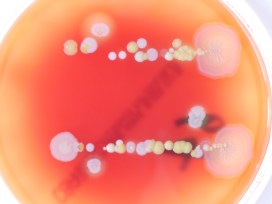
But let’s not get ahead of ourselves. Your body is full of bacteria. In fact, there are more bacteria than human cells in your body. Most of these bacteria are good for you; they help you digest food and protect you from diseases.

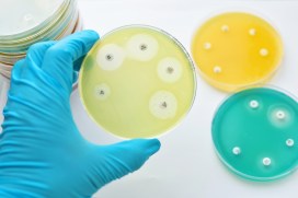
But once in a while a harmful bacterium causes an infection. This could be a lung, wound, or bladder infection, or something with a fancy name like, Black Death, tuberculosis, leprosy, syphilis or chlamydia. The doctor will then prescribe you antibiotics to kill the offending bacteria.

Hannah Behrens delivers her Science Short talk at the Museum

The development of antibiotics in the 20th century was a major breakthrough. For the first time bacterial infections could be effectively and rapidly treated. Since 1942, when antibiotics first became available, we have discovered many new antibiotics which have saved millions of lives.

However, in the last 30 years we have not managed to develop any new antibiotics. During the same time, many bacteria have adapted to become resistant to the antibiotics we do have. In 2017, a woman in the US died because she had an infection with bacteria that were resistant to all available antibiotics. It is estimated that already 700,000 people in Europe alone die because of resistant bacteria per year. What is happening?

Bacteria are forming a lawn on this plate (light areas); where an antibiotic has been spotted on the bacteria they die and leave the surface blank (dark areas).

Every time we treat bacteria with antibiotics, most die, yet a few resistant bacterial cells survive. Like Rudolph the red nosed reindeer, the resistant bacteria are usually at a disadvantage until a special situation arises (a foggy night for Rudolph; treatment with antibiotics for resistant bacteria).

Under usual circumstances, producing a resistance mechanism is a disadvantage: it wastes energy and slows down growth, so very few bacteria are resistant. Only when all the non-resistant bacteria are killed by antibiotics do the resistant ones thrive. They have no more competition, and have all the resources, such as food and space, to themselves.

The more we use antibiotics, the more resistant bacteria we get. It is essential not to use antibiotics carelessly.

More antibiotics are used in animal farming than on humans. If we eat less meat, and so reduce the farming of livestock for food, we may reduce the growth of resistance bacteria. Another approach is to only take antibiotics when the doctor prescribes them. Antibiotics do not help against viral infections like colds. In many low and middle income countries, antibiotics are available in supermarkets and it is no coincidence that these countries have higher levels of resistant bacteria.

The precision antibiotics research group in the Department of Biochemistry at the University of Oxford

Apart from avoiding the unnecessary use of antibiotics, scientists – including me – are trying to develop better therapies against bacteria. I study precision antibiotics: drugs that specifically kill one species of bacteria. The advantage of this is that all good bacteria remain unharmed and only the disease-causing species is targeted. This also means that only resistant bacteria from this one species get an advantage to thrive.

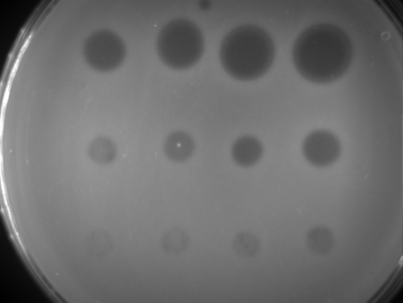
I am interested in species-specific antibiotics against Pseudomonas aeruginosa. This bacterial species causes lung and wound infections and, according to the World Health Organization, is one of the three bacteria for which we most urgently need new antibiotics. Colleagues of mine tested different precision antibiotics against Pseudomonas and found one that is better than the others, called Pyocin S5.

Hannah’s painting of how researchers think pyocin antibiotics kill bacteria. The pink bacterium produces pyocins (pink balls), which enter the susceptible blue bacteria through pores (blue). The blue bacteria mistake the antibiotic for a nutrient and open the pore to let it in. Once inside the bacterium it forms a pore in the inner membrane which causes leakage of the cell contents and kills the cell.

I am now investigating how stable this antibiotic is, how it recognises this specific species of bacteria and how it enters the bacterial cells. This knowledge is important to decide on how to store, transport and administer the drug. I also hope that understanding why Pyocin S5 is more effective than the other antibiotics will allow us to design more effective, targeted antibiotics in the future.

My hope is that one day we will treat all bacterial infections with precision antibiotics and that antibiotic resistance will become a problem of the past.

Bee beautiful

Our conservator Bethany Palumbo tells us how she restored a beautiful 19th-century papier-mâché model of a honeybee hive, created by master model-maker and anatomist Louis Thomas Jérôme Auzoux

Louis Thomas Jérôme Auzoux

Although the Museum’s collections are mostly of organic specimens, we also hold a fascinating collection of scientific models made to represent the natural world, made from all types of materials, from wax and cardboard to plaster and paint.

We are lucky enough to own a model made by esteemed French anatomist Louis Auzoux (1797-1880), who in the late 19th century developed a method of building strong yet light papier-mâché models that could be taken apart and rebuilt, allowing internal elements such as tissues and organs to be studied in detail.

Model of a honeybee hive in box with six bees, by Louis Auzoux

While Auzoux made many models demonstrating human anatomy, he later expanded his business to include magnified models of plants and insects. The model we have is of a honeybee hive, containing six beautiful bees.

The hive, painted with a protein-based paint and varnished with gelatine, is large enough to allow the viewer to see the fine details of the hive, including individual chambers containing tiny larvae.

As you can see in the image at the top of the article, the bees themselves are also intricately painted, with rabbit hair used to simulate their natural fuzz, and delicate wings constructed from metal wire.

While there was much to admire about this model, it was in received in poor condition. Previous restoration attempts had introduced many materials that were now failing. There were fills, constructed of paper, applied to areas in an attempt to hide cracks in the original model. These were covered in oil paint, which was dripping over the original paintwork and had become brittle and discoloured.

Oil paint layers were peeling from the model

The whole hive was coated in a layer of cellulose nitrate film, a popular coating in the mid-20th century which was used as protection and to create a gloss finish. This coating doesn’t age well, resulting in peeling. It had also been applied to the bees themselves, clumping together the bee ‘fuzz’ and disguising the paintwork underneath.

The priority for treatment was to return the model to its original form while stabilising it for the future.

I undertook treatment in several stages over the course of six months. First, the cellulose nitrate film was removed from all areas using acetone, which could be applied with a cotton bud and fortunately didn’t affect the paint layer beneath.

Fill material used to cover previous damage had become discoloured

The next stage was to remove the discoloured oil paint from the hive. This was done manually using metal and wooden tools lubricated with white spirit, which were used to gently scrape the surface under magnification. This revealed old fills on the hive, made from a combination of plastic tape, paper and old adhesives which also needed to be removed. They were easily softened with water and gently peeled away.

Once all unstable introduced materials were removed, work began to stabilise the original model. The bees were suffering from paint cracking and peeling, as seen in the magnified photograph below.

Peeling paint at 6x magnification

We decided to consolidate this using gelatine as it would be in keeping with the original construction and could easily be reversed if necessary. Gelatine was mixed in water and warmed to make it a thin consistency, and then applied with a paintbrush. Once the paint flakes had softened they could be gently pressed down. Gelatine was also used with acid-free tissue to stabilise the cracks and areas of surface loss on the hive.

With the hive and bees now clean and stable, the quality of this piece and its incredible paintwork can really be admired. We hope to put it on display soon for all our visitors to enjoy!

Crafty camouflage

Last week we brought you snails that attach all manner of pebbles, fossils, corals and shark teeth to their shells. Today we give you a newly-discovered fossil green lacewing larva that attached pieces of soil to its body as an act of camouflage. Our research fellow Ricardo Pérez-de la Fuente, lead author of the new paper, explains…

Visual camouflage is one of the most successful survival strategies in nature. Camouflaging is usually defensive, allowing animals to be left unnoticed by their predators, but it can also be used aggressively by predators themselves to approach their prey undetected.

Some camouflaging animals can actively change their colouring to match that of the background ‒ a technique called crypsis. Others can make their bodies resemble elements of the environment, such as leaves or twigs, which is called mimicry.

Italochrysa italica, an extant green lacewing larva carrying a dense debris packet made of soil fragments. Taken from the open access publication Tauber & Winterton, 2014.

Yet another approach to camouflage involves collecting diverse materials from the environment and incorporating them on the animals’ bodies in order to better blend with the surroundings. This is known as debris-carrying, trash-carrying, or decoration, and it can be found across a wide variety of animals including sea urchins, gastropods, and arthropods, such as decorating crabs, or sand- and mud-covering spiders.

My colleagues and I have just published the discovery of a fossil green lacewing larva, pictured at the top of the article, that has been preserved carrying bits of soil that it used for camouflage and physical protection. It’s a new larval species just 1.5 mm in length, and is preserved in Early Cretaceous Lebanese amber. We have named it Tyruschrysa melqart after the Phoenician city of Tyre and its tutelary god Milk-Qart (if you want to learn the reasons behind this name check out our open access paper!).

Interpretative drawing of Tyruschrysa melqart: body in grey, ‘tubes’ with setae coloured according to which body part they are attached to, and soil debris in brown.

Green lacewing larvae are active predators that eat other insects such as aphids, using sickle-shaped ‘jaws’ to pierce their prey, suck out their fluids and liquefy their tissues; eating is easier when there is no need to chew! Some green lacewing larvae are debris carriers, entangling all kinds of debris among their velcro-like ‘hairs’ called setae, which extend from relatively short ‘bumps’ on their backs. This debris is carefully selected and gathered with meticulous head and body movements to form a so-called debris packet on the back of the insect.

‘Tubes’ bearing setae of Tyruschrysa melqart, with detail of their mushroom-shaped endings (bottom), used for anchoring bits of soil.

The new fossil and similar ones described from younger Cretaceous ambers differ from modern relatives because instead of short ‘bumps’ with setae on their backs they have relatively long ‘tubes’, giving them a bizarre appearance.

These tubes have setae with mushroom-shaped endings of a kind never seen before in extinct or living green lacewing larva species. The mushroom-shaped ending is a special adaptation to anchor debris, which in the case of Tyruschrysa melqart are fragments of soil.

Hallucinochrysa diogenesi, another Cretaceous green lacewing larva bearing long ‘tubes’ with setae on its back, but carrying a debris packet made of plant hairs (trichomes). Preserved in Spanish amber (105 million years old).

It was already known that Cretaceous green lacewing larvae like Tyruschrysa had long tubes on their backs and that they collected plant hairs and other plant material to construct their packet of debris. But thanks to the new discovery we now know that these immature insects also used bits of soil, and that in the deep past debris packets were probably as diverse as those we see today.

Green lacewing larvae have been gathering debris to camouflage and protect themselves for about 130 million years, giving rise to the different body adaptations we see amongst these fascinating tiny collectors.

‘A soil-carrying lacewing larva in Early Cretaceous Lebanese amber’ Ricardo Pérez-de la Fuente, Enrique Peñalver, Dany Azar and Michael S. Engel is published as open access in Scientific Reports this month.

Bacteria: captured and cultured

For our new exhibition, Bacterial World, we embarked an exciting science/art experiment to make visible the colonies of bacteria present on a wide range of our everyday items and belongings. Once cultured and photographed, eight of these colonies were captured by artist Elin Thomas as a set of crochet artworks that are on display in the exhibition. Our exhibitions officer Kelly Richards tells us more…

For every human cell in your body, a bacterial cell is also present. These bacteria are part of our microbiome, a vast array of microorganisms that use our body as a home and our food as a source of nutrients. In return, the bacteria help us to digest food, maintain our immune systems and keep dangerous bacteria at bay. In fact without these bacteria we would be very sick indeed.

It’s hard to see our microbiome because individual bacteria can easily be as small as 0.2 microns; you could fit over a thousand of these smallest bacteria on one side of a red blood cell. But if we can select and artificially grow the bacteria, their colonies become living, breathing cities visible to the naked eye.

Click the images above to find out more about culturing bacterial colonies

Colonies, both natural and artificial, can contain billions of bacteria as well as the materials that they secrete such as slime, which helps them to move across surfaces, and antibiotics, which kill off other bacterial colonies that could compete for food and space. In their attempts to dominate the space and food available, as well as get enough oxygen to live, colonies can create beautiful, complex structures.

We had a go at visualising the bacteria that live invisibly alongside us by asking visitors to take part in a simple experiment. With the help of microbiologist Rachael Wilkinson, we took items such as coins, keys and jewellery and touched them lightly against agar plates – dishes containing a nutrient-rich jelly that aids bacterial growth. The agar plates were then given to Nicole Stoesser, a clinical microbiologist at the John Radcliffe Hospital, who grew them in the safe environment of the laboratory.

Many different types of colonies grew from the objects we printed. In the collage above, eight of these colonies have been represented as crocheted Petri dishes by artist Elin Thomas. These artworks are on display in the Bacterial World exhibition until 28 May 2019.

In the gallery below is a photograph of every participant’s plate, whether anything grew in it or not. Click on an image to see a larger version. If you took part in the experiment you will be able to identify your own plate from its number.

The results go to show that we really are living in a bacterial world!

Restoring the Great Lizard of Stonesfield

by Paul Smith, Museum director

One of the unusual things about the collections in the Museum is that some of the specimens date back hundreds of years, and so have been researched by generation after generation. Sometimes these specimens have been damaged and repaired, and in some cases this has happened many times, leaving a complex history of research and conservation.

One high profile example is the type specimen of the theropod dinosaur Megalosaurus bucklandii – the world’s first scientifically described dinosaur. This specimen itself is the lower jaw, pictured above, which has been in the collections of Oxford University since 1797 at the latest.

Working with the Centre for Imaging, Metrology, and Additive Technology (CiMAT) at WMG, University of Warwick, we have been unraveling the conservation and repair history of the fossilised jaw using an innovative combination of modern technologies.

Identification of repair using X-ray computed tomography (XCT ) from the Megalosaurus bucklandii type specimen. The two colours indicate two different types of plaster material. Scale bar is 10 cm.

Earlier studies had mapped the presence of plaster used for repair, but X-ray CT scanning of the type used in medical procedures rapidly revealed a number of different phases of repair. In each of these repairs the plaster was of different composition and was used in different places.

One type, shown in red on the image above, was used to infill fractures and to make the specimen more robust; a second type, shown in green, was used to repair the teeth and, in some cases, to recreate the teeth. Interestingly, the extent of plaster revealed by the CT scanning is actually less than previously interpreted with the naked eye.

The two types of plaster were then analysed chemically to better understand their historical use, revealing quite different compositions. The more abundant ‘red’ plaster is mixed with quartz sand and calcite grains, possibly from the rock matrix surrounding the fossil, to make it look more similar.

Carbon is also abundant and grains rich in lead are present. Carbon is not common in the rock itself, and the carbon in the plaster has probably come from a varnish such as shellac being used to coat the repair.  The presence of lead was more puzzling. Further analysis eventually showed that the grains were made of red lead – lead tetroxide – which was used historically as a pigment in paint. The red lead in the repair may have been used to colour the plaster, but it may also have been applied to make the density of the plaster more similar to that of the fossilized bone, and so replicate the weight of the specimen better.

Reconstruction of Megalosaurus bucklandii by Julius T. Csotonyi

The second type of plaster, used to repair the teeth, lacks the lead of the first type but contains barium. Barium hydroxide was often used as a consolidant and sealant for plaster, which would explain its use here.

Having a full understanding of the repair history and the position and extent of plaster helps us in a number of ways. It allows researchers to understand which parts of the lower jaw are original and anatomically reliable, and it helps the curators and conservators to know which parts of the specimen are more fragile during handling and display.

By combining cutting-edge scanning technologies with heritage material we are able to shed new light on the conservation history, and future, of important specimens such as Megalosaurus bucklandii.

Read the full paper here.

Marvellous Mantodea

In the latest display in our Presenting… series, collections manager Amoret Spooner takes a look at the wonderful and sometimes strange world of the praying mantis.

Praying mantis is the common name given to an order of insects called Mantodea, a word which derives from mantis meaning prophet, and eidos meaning form or type. The more familiar ‘praying mantis’ refers to the striking way that they hold their large forelimbs, in a ‘praying’ posture.

Display of different mantis species

There are over 2,400 species of mantis worldwide, split into 21 different families. The image above shows their incredible diversity of colour, shape and size. But while they may differ in appearance, their biology and many behavioural traits are the same.

Mantis are predators of insects, including other mantis, but larger species will eat small lizards and birds. But they are perhaps best known for being cannibalistic. This behaviour is most commonly seen in nymphs straight out of the egg case, or ootheca, but it can also occur when the female eats the male after mating. However, cannibalism is not required to mate, so when it happens it’s usually because the female is hungry!

The egg case, or ootheca, of mantis vary greatly depending on the size and behaviour of the species.
Revisio Insectorum Familiae Mantidarum was one of John Obadiah Westwood’s greatest works. Thankfully he kept all of his drawings, annotated pages and notes for the publication, allowing us an insight into the years of work he put into its production.

Praying mantis are ambush hunters, either camouflaging themselves while waiting for their prey to approach, or actively stalking prey. Their compound eyes are specialised in perceiving motion, and are widely spaced giving them a wide field of vision. Along with powerful front legs and an ability to move the head up to 180°, this makes them successful predators.

The Museum’s archive contains original drawings and annotations by John Obadiah Westwood (1805–1893), the first Hope Professor of Zoology. As a renowned scientist Westwood described many new mantis species, and he was also a talented artist.

The Presenting… Marvellous Mantodea case is on display at the Museum until 1 November 2018.