One of the unusual things about the collections in the Museum is that some of the specimens date back hundreds of years, and so have been researched by generation after generation. Sometimes these specimens have been damaged and repaired, and in some cases this has happened many times, leaving a complex history of research and conservation.
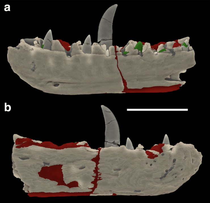
One high profile example is the type specimen of the theropod dinosaur Megalosaurus bucklandii – the world’s first scientifically described dinosaur. This specimen itself is the lower jaw, pictured above, which has been in the collections of Oxford University since 1797 at the latest.
Working with the Centre for Imaging, Metrology, and Additive Technology (CiMAT) at WMG, University of Warwick, we have been unraveling the conservation and repair history of the fossilised jaw using an innovative combination of modern technologies.
Identification of repair using X-ray computed tomography (XCT ) from the Megalosaurus bucklandii type specimen. The two colours indicate two different types of plaster material. Scale bar is 10 cm.
Earlier studies had mapped the presence of plaster used for repair, but X-ray CT scanning of the type used in medical procedures rapidly revealed a number of different phases of repair. In each of these repairs the plaster was of different composition and was used in different places.
One type, shown in red on the image above, was used to infill fractures and to make the specimen more robust; a second type, shown in green, was used to repair the teeth and, in some cases, to recreate the teeth. Interestingly, the extent of plaster revealed by the CT scanning is actually less than previously interpreted with the naked eye.
The two types of plaster were then analysed chemically to better understand their historical use, revealing quite different compositions. The more abundant ‘red’ plaster is mixed with quartz sand and calcite grains, possibly from the rock matrix surrounding the fossil, to make it look more similar.
Carbon is also abundant and grains rich in lead are present. Carbon is not common in the rock itself, and the carbon in the plaster has probably come from a varnish such as shellac being used to coat the repair. The presence of lead was more puzzling. Further analysis eventually showed that the grains were made of red lead – lead tetroxide – which was used historically as a pigment in paint. The red lead in the repair may have been used to colour the plaster, but it may also have been applied to make the density of the plaster more similar to that of the fossilized bone, and so replicate the weight of the specimen better.
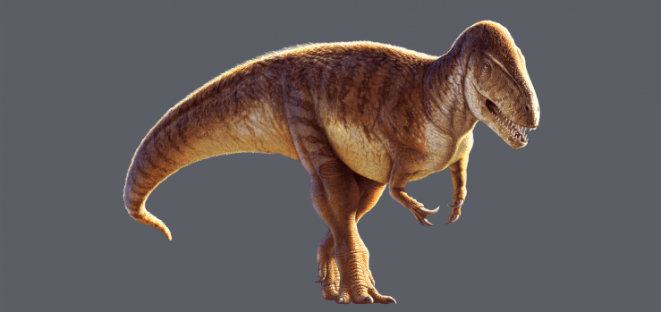
Reconstruction of Megalosaurus bucklandii by Julius T. Csotonyi
The second type of plaster, used to repair the teeth, lacks the lead of the first type but contains barium. Barium hydroxide was often used as a consolidant and sealant for plaster, which would explain its use here.
Having a full understanding of the repair history and the position and extent of plaster helps us in a number of ways. It allows researchers to understand which parts of the lower jaw are original and anatomically reliable, and it helps the curators and conservators to know which parts of the specimen are more fragile during handling and display.
By combining cutting-edge scanning technologies with heritage material we are able to shed new light on the conservation history, and future, of important specimens such as Megalosaurus bucklandii.
In the latest display in our Presenting… series, collections manager Amoret Spooner takes a look at the wonderful and sometimes strange world of the praying mantis.
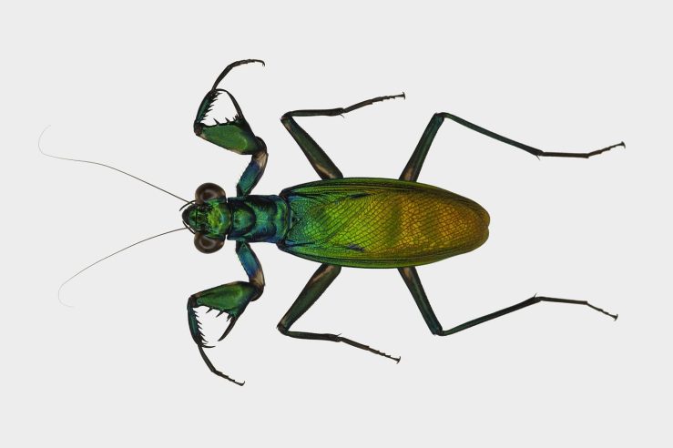
Praying mantis is the common name given to an order of insects called Mantodea, a word which derives from mantis meaning prophet, and eidos meaning form or type. The more familiar ‘praying mantis’ refers to the striking way that they hold their large forelimbs, in a ‘praying’ posture.
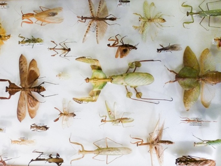
There are over 2,400 species of mantis worldwide, split into 21 different families. The image above shows their incredible diversity of colour, shape and size. But while they may differ in appearance, their biology and many behavioural traits are the same.
Mantis are predators of insects, including other mantis, but larger species will eat small lizards and birds. But they are perhaps best known for being cannibalistic. This behaviour is most commonly seen in nymphs straight out of the egg case, or ootheca, but it can also occur when the female eats the male after mating. However, cannibalism is not required to mate, so when it happens it’s usually because the female is hungry!
The egg case, or ootheca, of mantis vary greatly depending on the size and behaviour of the species.Revisio Insectorum Familiae Mantidarum was one of John Obadiah Westwood’s greatest works. Thankfully he kept all of his drawings, annotated pages and notes for the publication, allowing us an insight into the years of work he put into its production.
Praying mantis are ambush hunters, either camouflaging themselves while waiting for their prey to approach, or actively stalking prey. Their compound eyes are specialised in perceiving motion, and are widely spaced giving them a wide field of vision. Along with powerful front legs and an ability to move the head up to 180°, this makes them successful predators.
The Museum’s archive contains original drawings and annotations by John Obadiah Westwood (1805–1893), the first Hope Professor of Zoology. As a renowned scientist Westwood described many new mantis species, and he was also a talented artist.
The Presenting… Marvellous Mantodea case is on display at the Museum until 1 November 2018.
This article is taken from European research magazine Horizon as part of our partnership to share natural environment science stories with readers of More than a Dodo. For more on the development of the brain see our Brain Diaries exhibition site.
One of the major features that distinguishes humans from other primates is the size of our brains, which underwent rapid evolution from about two to three million years ago in a group of our ancestors in Africa called the Australopithecines. During this period, the human brain grew almost three-fold to reach its current size. Scientists know this from skull remains, but have puzzled over how it happened…
This year, the mystery was partially solved by Professor Pierre Vanderhaeghen at the Flanders Institute for Biotechnology in Belgium. Prof. Vanderhaeghen, who was conducting his work as part of the GENDEVOCORTEX project, went on a hunt for the genes that drove the growth of human brains.
Scientists had suspected that brain expansion began in our human ancestors when they evolved genes that are switched on in the foetus, when a lot of key brain development occurs. Prof. Vanderhaeghen therefore looked for genes present in human foetal tissue, but missing from our closest living relatives, apes.
His lab discovered 35 hominid – present only in apes and humans – genes that were active in foetal brain tissue. They then became intrigued by three specific genes – all similar to NOTCH genes, an ancient gene family involved in sending messages between cells and that are present in all animals. They found that the three new genes, collectively named NOTCH 2NL, were created by a “copy and paste error” of an original NOTCH gene.
This error created entirely new proteins which likely helped our ancestors’ cerebral cortex to balloon. This is the part of our brain responsible for our language, imagination and problem-solving abilities. Scientists at the University of California, Santa Cruz, have also identified the NOTCH 2NL genes in DNA from Homo sapiens’ extinct cousins – the Neanderthals and Denisovans.
(The NOTCH 2NL) genes are only present in humans today. They were also present in Neanderthal DNA, but not in chimpanzees
Prof. Vanderhaeghen
Evolution These genes control the growth rate and differentiation of brain stem cells – the starter cells that multiply and give rise to all neurons in our brain – causing them to seed more nerve cells, which in turn helped to expand brain size. The genes likely led to more neurons and brain tissue in our ancestor’s descendants – including Neanderthals, Denisovans, and modern humans.
Prof. Vanderhaeghen’s research could also help to provide new insights into brain disorders. The US researchers linked genetic faults in DNA that were very similar to NOTCH 2NL, to children born with enlarged brains or small brains. Many of the new human-specific genes are located in a small area of our genome that plays an important role in brain size, according to Prof. Vanderhaeghen.
As DNA in this area closely resembles another part of the genome where it was originally cut and pasted from millions of years ago, errors are more likely, said Prof. Vanderhaeghen. “Patients who have (inherited) deletions in this area tend to be at risk of developing schizophrenia, whereas patients with duplications are more at risk of autistic spectrum disorder,” he said.
Prof. Vanderhaeghen is now studying some 20 of the remaining human-only genes to see how they contributed to the evolution of the human brain.
Something like 40-50% of the Neanderthal genome can still be found in people today.
Prof. Svante Pääbo, Max Planck Institute for Evolutionary Anthropology, Leipzig, Germany
The use of genetics to study human evolution in this way is helping to transform our understanding of how our own species compared to our ancestors. Traditionally, scientists have studied extinct species by looking at the fossilised remains of their bones. This was how they discovered the existence of Neanderthals, the extinct human species that lived across Europe and much of Asia before vanishing around 40,000 years ago.
In the last decade, however, scientists have begun to look at the DNA inside these bones. Professor Svante Pääbo, director of the Max Planck Institute for Evolutionary Anthropology in Leipzig, Germany, has led the way in sequencing DNA of these extinct humans from small bone fragments.
This allows scientists to compare modern human DNA with that of extinct humans, rather than just living relatives like chimps. Already, the work has revealed some surprising findings – our own species appears to have interbred with some of these ancient relatives during our history.
Ancient humans Scientists have found that the DNA of every person outside Africa is 1-2% Neanderthal, meaning that these extinct human relatives had offspring with our own ancestors.
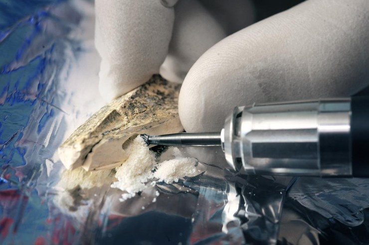
An international consortium of researchers is sequencing the 3 billion bases that make up the genome of our closest relative – the Neanderthal. The sequence is generated from DNA extracted from three Croatian Neanderthal fossils using novel methods developed for this project. Image credit – Frank Vinken for Max Planck Society
“Different people tend to carry different pieces of the Neanderthal genome,” said Prof. Pääbo, who is undertaking a project called 100 Archaic Genomes to decipher the DNA of ancient human individuals. “Something like 40-50% of the Neanderthal genome can still be found in people today,” he said.
According to Prof. Pääbo, we retained some of this DNA because it offered an advantage to our ancestors. “Some (of this retained DNA) has to do with the immune system, presumably helping us to fight off infectious diseases.”
The power of genetics to unravel the history of human evolution took a new twist in 2010 after Prof. Pääbo’s lab sequenced DNA from a finger bone fragment found by a Russian archaeological team in a remote Siberian cave.
The analysis revealed the bone belonged to a previously unknown human relative, now called Denisovans after Denisova Cave where the bone was found. This mysterious ancient human species lived at around the same time as Neanderthals, but further east into Asia.
Last year, Prof. Pääbo’s group published DNA sequences from a tooth found in the cave – the fourth ever Denisovan discovered. We now know Denisovan DNA carries more variation than Neanderthal DNA, leading scientists to conclude that they were more widespread than the better-known Neanderthals.
Denisovans left a more impressive stamp on some of us than Neanderthals, according to Prof Pääbo. Their DNA can be found in people across Asia today, while indigenous peoples of Papua New Guinea and Australia may carry up to 5%. Tibetans also carry some Denisovan DNA in their genomes, which has helped them adapt to life at high altitudes where there is little oxygen in the atmosphere.
Prof. Pääbo and his colleagues will soon publish their third high-quality genome – where almost the entire DNA sequence is intact – of a Neanderthal from Siberia. A deciphered genome of this quality allows for better DNA comparisons and could tell us more about the evolution of important genes – such as those linked to the development and function of the brain. It will add yet another puzzle piece to help us understand the history of our closest extinct relatives, according to Prof. Pääbo.
“There may even be other forms of extinct humans out there to be discovered by studying the DNA of the (ancient) bones we find,” he said.
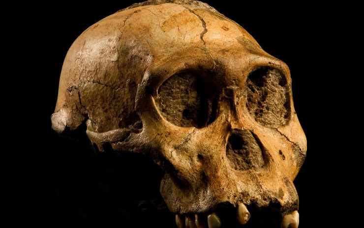
Top image: The skull of a Australopithecus sediba, a species of Australopithecines, who were our ancestors and whose brains started to grow two to three million years ago. Image credit – Australopithecus sediba by Brett Eloff, courtesy Profberger and Wits University is licensed under CC BY-SA 4.0.
Cephalopods are a remarkable group of molluscs that includes nautilus, octopuses, cuttlefish and various groups of ‘squid’. The other major groups of molluscs includes more familiar shelled animals such as gastropods (snails and slugs), bivalves, and chitons, as well as some less familiar forms.
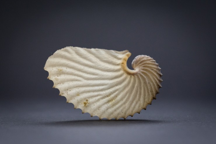
In natural history museums, molluscs are normally represented by shell collections because the hard shelly parts are easier to preserve and store than the soft tissue. This creates a bias against the soft-bodied cephalopods, such as squids, octopuses and cuttlefish, because aside from the cuttlebones of cuttlefish and the thin gladius in squids there aren’t many hard parts that can be preserved to represent these animals in dry collections. For octopuses it’s normally only the beak and microscopic radulae, a toothed tongue-like structure, that can be preserved. But there is one notable exception: the eggcases of argonauts.
Model of Argonauta argo. Image: Mark Carnall
Argonauts, four* species of octopuses in the genus Argonauta, are unusual in that they produce a paper-thin eggcase, sometimes referred to as a shell. Unlike a true shell it’s not attached to the body of the argonaut, but secreted by two specialised webbed arms. The eggcases themselves are sometimes called paper nautiluses because they resemble the spiral shells of nautiluses, but they are structurally and functionally very different.
External morphology of a female paper nautilus (Argonauta argo) with eggcase. Poli, Giuseppe Saverio. Testacea utriusque Siciliae. (1791-1827). http://biodiversitylibrary.org/page/44020354
Argonaut eggcases wash on up shorelines around the world and have been known for centuries. But it’s only comparatively recently that the origin and use of these cases has been described. When eggcases containing live argonauts were first encountered it was supposed that argonauts were reusing empty shells created by another animal, much like hermit crabs repurpose gastropod shells.
Pioneering research by marine biologist Jeaneatte Villepreux-Power in the 19th century led to observations of Octopus and Argonauta, confirming that the eggcases are made and repaired by female argonauts. It wasn’t until 2010 that we understood how argonauts use these cases to float in the ocean. It turns out that they ‘bob’ their shells to gulp a pocket of air. Then, using their second pair of arms, they trap the air in the top of the shell and dive releasing enough air to maintain the required buoyancy.
Only female argonauts make the eggcases, so the free-floating males are tiny in comparison. In addition to providing a home for female argonauts, these structures are used to brood embryos in. One eggcase was described with nearly 50,000 embryos attached to the inside of the shell.
Preparation showing series of argonaut egg cases of varying sizes.
Thanks to their oddity and beauty these eggcases are common in museum collections, but they represent one of the marvels of evolution. Unlike many bottom-dwelling octopuses, female argonauts have evolved this amazing structure to function as an underwater craft to allow them to leave the ocean floor and inhabit the open oceans: the true astronauts of the sea.
To celebrate the pioneering work of Jeaneatte Villepreux-Power, these amazing animals, their eggcases, and a selection of museum specimens are on display in the Museum’s Presenting… case until the 3 July 2018.
You may have heard of the Cambrian Explosion, an ‘event’, starting roughly 540 million years ago, when all the major animal groups suddenly appear in the fossil record, an apparent explosion of life and evolution.
But was there really an evolutionary explosion of all these animal groups, or is the lack of evidence from earlier periods due to some peculiarity of the fossilisation process? The debate has rumbled on for a number of years.
Now, a new study from our research team, the University of Oxford’s Department of Zoology, and the University of Lausanne, claims that the early Cambrian saw the origins and evolution of the largest and most important animal group on Earth – the euarthropods – in a paper which challenges two major pictures of animal evolution.
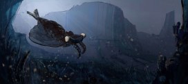
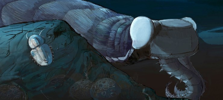
Euarthropoda contains the insects, crustaceans, spiders, trilobites, and a huge diversity of other forms alive and extinct. They comprise over 80 percent of all animal species on the planet and are key components of all of Earth’s ecosystems, making them the most important group since the dawn of animals over 500 million years ago.
Exceptionally preserved soft-bodied fossils of the Cambrian predator and stem-lineage euarthropod Anomalocaris canadensis from the Burgess Shale, Canada. Top left: Frontal appendage showing segmentation similar to modern-day euarthropods. Bottom right: Full body specimen showing one pair of frontal appendages (white arrows) and mouthparts consisting of plates with teeth (black arrow) on the head. Images: A. Daley.
A team based at the museum, and now at Lausanne, conducted the most comprehensive fossil analysis ever undertaken on early euarthropods, to try and establish whether these animals really did emerge in the early Cambrian period, or whether fossilisation just didn’t occur in any earlier periods.
In an article published today in the Proceedings of the National Academy of Sciences they show that, taken together, the total fossil record does show a gradual radiation of euarthropods during the early Cambrian, 540-500 million years ago, challenging other ideas that suggest either a rapid explosion of forms, or a much slower evolution that has not been preserved in the fossil record.
Each of the major types of fossil evidence has its limitation and they are incomplete in different ways, but when taken together they are mutually illuminating Professor Allison Daley
Reconstruction of the Cambrian predator and stem-lineage euarthropod Anomalocaris canadensis, based on fossils from the Burgess Shale, Canada. Reconstruction by Natalia Patkiewicz.
By looking at a huge range of fossil material the researchers ruled out the possibility that Pre-Cambrian rocks older than around 541 million years would not have preserved early euarthropods. The only plausible explanation left is that the origins of this huge animal group didn’t evolve until about 540 million years ago, an estimate which also matches the most recent molecular dating.
The timing of the origin of Euarthropoda is very important as it affects how we view and interpret the evolution of the group and its effects on the planet. By working out which groups developed first we can trace the evolution of physical characteristics, such as limbs.
Exploring all the evidence like this allows us to make an informed estimate about the origins of key animal groups, leading to a better understanding of the evolution of early life on Earth.
Model of the Cambrian stem lineage euarthropod Peytoia, based on fossils from the Burgess Shale. Top left: Closeup of the mouth parts and frontal appendages. Bottom right: Overall view of the body. Model and image: E. Horn.
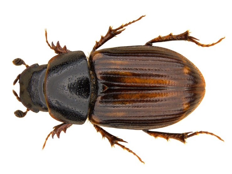
In a previous article on this blog I reported the discovery, in an insect collection, of the 21st British specimen of the ‘Regionally Extinct’ dung beetle Melinopterus punctatosulcatus. And since then, I’ve been on the hunt for more…
Heading out to numerous other museum collections I discovered more specimens, all collected in the same locality – Deal in Kent. In Ipswich Museum there are six, collected by C. Morley in 1896; there are two in the Natural History Museum, London, collected by G.C. Champion; and in the Museum of Zoology, Cambridge there are two collected by G.C. Hall in 1883.
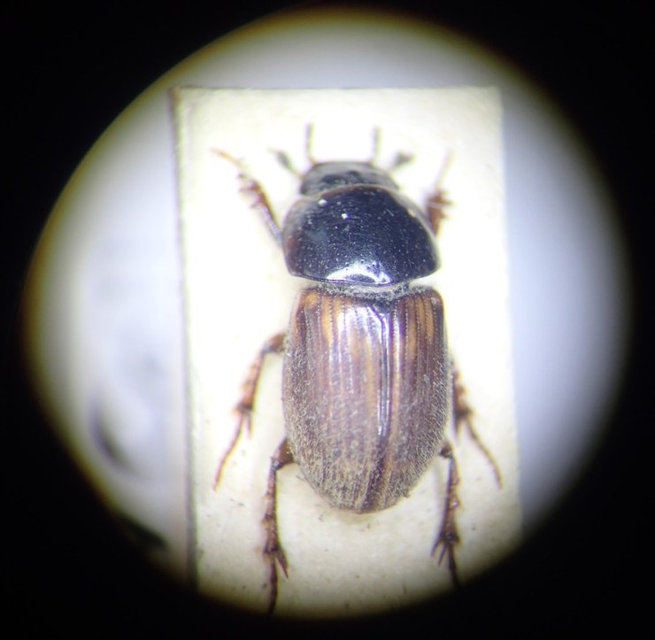
A view through the microscope of Melinopterus punctatosulcatus held in the collections of Ipswich Museum, collected by C. Morley in 1896
But the earliest and most recent finds are both in the National Museum of Scotland – one from May 1871, in the G.R. Waterhouse collection, and one from 1923, in the T. Hudson-Beare collection. So now we know of 42 specimens of this beetle with data and we know that the species occurred at Deal for about 50 years. But why are there no records after this time?
The Deal sandhills in Kent were famous for their insects, but even as long ago as 1900 entomologists* were discussing the negative impact of “summer camping-out stations and the modern craze for the ‘Royal and Ancient Game of Golf'” on beetles and butterflies in the area.
Today, most of the sandhills are gone and there are no grazing animals other than a few rabbits. Most of the surrounding land is either developed as a golf course or under agricultural management. So, is the possible local extinction of this dung beetle due to habitat loss and a lack of dung?
Deal, Kent: the original locality for Melinopterus punctatosulcatus, with remnants of the sandhills in the distance
To try and answer this question, naturally I went looking for poop in Deal. In a field in Sandwich Bay I could hear sheep bleating in the distance, although poo was scarce. Eventually I found a few old plops and inside were ten Calamosternus granarius, a small dung beetle. This was good, but my main target was Melinopterus punctatosulcatus.
A specimen from the Museum of Melinopterus punctatosulcatus, previously listed as ‘Regionally Extinct’ in Britain, but now rediscovered in Deal, Kent
I probed the poop further. To my delight, crowded in the remaining squishy bit were four other species. On close inspection, one of these was hairy, so a male, and much darker than its close relatives. It fitted perfectly with my expectations for Melinopterus punctatosulcatus after seeing so many examples in museum collections. Success! This beetle, misidentified in museum collections for so long, and not seen since the 1920s in Deal, is indeed hanging on in Kent.
Disappointingly, after a further few days of searching, only a handful more specimens were seen. This suggests that either the species exists at low population levels, or that it was it was not the peak emergence period when I was there. Nonetheless, a species not recorded anywhere in the UK for over 70 years is actually still here.