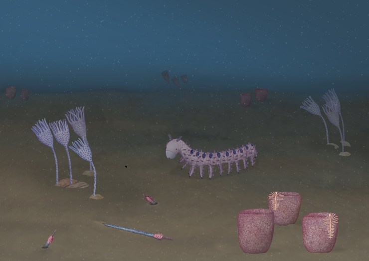
Abigail Harris - artwork showing reconstruction of Cambrian ocean animal life

Cambrian creation

Abigail Harris - artwork showing reconstruction of Cambrian ocean animal life

by Abigail Harris

Over the past few months our researchers have been working with University of Plymouth illustration student Abigail Harris, who has delved into the weird and wonderful world of some of the earliest animals. Here, Abigail tells us about the process that led to the creation of her Cambrian artwork, inspired by our First Animals exhibition.

I first visited the Museum in April this year when I was given the opportunity to collaborate with scientists as part of a module in my BA in at the University of Plymouth. Things kicked off with a short talk about the Ediacaran and Cambrian geological periods, when Earth’s first animal life started to appear.

I quickly narrowed my interest down to fossils from the Cambrian period which are more complex life forms, more similar to life today. A collection of small fossils from the Chengjiang fossil site in Yunnan province, China was the inspiration for some initial observational drawings.
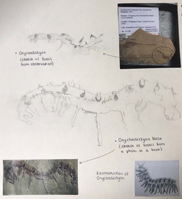
Abigail Harris - sketches for artwork showing reconstruction of Cambrian ocean animal life
A sketchbook page showing initial sketches and observations of Onychodictyon
Final illustration of Cotyledion

After returning to Plymouth University, I began to develop these initial sketches and observations, continuing to research the Chengjiang material and learning more about the characteristics of some of the creatures preserved as fossils.

I wanted to create an under-the-sea ecology reconstruction showing a diversity of life forms, focusing on Onychodictyon, Cotyledion, Cricocosmia, Luolishania, and Paradiagoniella.

A five-step process was used for each reconstruction. Initially, I would sketch the fossil as I saw it, then I would research the characteristics and features of that animal, making a list of things to include in my drawing. A second drawing would then include all of these characteristics, not just what was initially visible in the fossil.

These rough sketches were then sent to the scientists for feedback, helping me to redraw and paint the illustrations with watercolour, before scanning and digitally editing each painting. Lastly, I created a background and added my illustrations.

Initial under under the sea ecology reconstruction.

Although the reconstructions were not completely finished by the time of my project deadline, I returned to the Museum in July and was given a tour of the First Animals exhibition by Deputy Head of Research Imran Rahman, as well as the opportunity to discuss how to improve my artworks for accuracy.

Another round of sketching and painting led to the final piece, shown at the start of this article, complete with an added digital background of the seafloor, and darkened to reflect the murky world of a Cambrian ocean, 50 metres below the surface.

Tests of time: Foraminifera and Radiolarians in science, art and 3D

Doctoral researcher Elaine Charwat is exploring the value and meaning of models and casts in the Museum’s collections as part of her PhD. She has recently been studying some fabulous models that help to visualise and understand some of the very, very smallest of specimens…

By Elaine Charwat

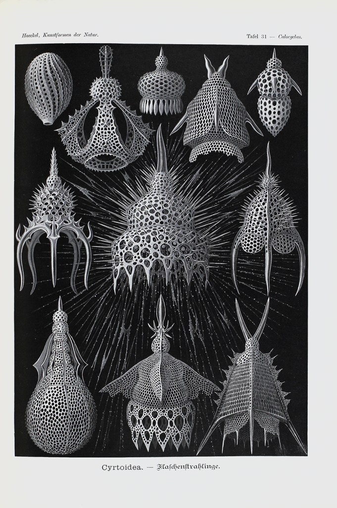
The first time I encountered a Radiolarian was in a book – Ernst Haeckel’s (1834-1919) weird and wonderful Kunstformen der Natur (Art Forms in Nature, 1899-1904). It took comparative morphology – comparing the shapes of organisms – to new giddy heights, scientifically, philosophically and artistically. I felt that giddiness when looking at page after page crammed with crustaceans, orchids, hummingbirds, moths and even bat faces, all exquisitely arranged to celebrate their symmetries, the evolution and kinship of their shapes and forms. It also made visible organisms that are normally all but invisible.

Illustration of Cyrtoidea (table 31) from Kunsterformen der Natur (1899 – 1904) by Ernst Haeckel. By permission of the Linnean Society of London.

Foraminifera and Radiolarians are microscopic sea-dwelling organisms. Species may be found as fossils dating from Cambrian times, ca. 500 million years ago, right up to living specimens today.

To Haeckel, they were living proof of Darwin’s theory of evolution, and for his own belief that morphology was the key to understand the actual processes of evolution, catching it in the act. However, these organisms had two big disadvantages – their unwieldy taxonomy, or the way they are classified, and their minute size: they were difficult to examine and display.

Illustrations of Radiolarians, (table 28). from Die Radiolarien (1862) by Ernst Haeckel. By permission of the Linnean Society of London.

Through his illustrations, Haeckel widely popularized them – triggering a Victorian craze for microscopes and microorganisms, as well as influencing art nouveau art and architecture. But there were limits to what an illustration could communicate. Models stepped in, representing these organisms in ways illustrations could not.

Detail from Haeckel’s Kunstformen der Natur (1899 – 1904)

One defining feature of Radiolarians and Foraminifera is their shells – called “tests”. Variations in shapes of the tests not only indicate that they are different species, but also, excitingly, provide clues about space and time. The tests of Neogloboquadrina pachyderma, for instance, record ocean temperature over geological timescales – their shells coil to the left when water temperatures are relatively cold, and to the right when it is warmer. The potential for research into climate change is obvious. Foraminifera are also important “signature fossils”, helping geologists to determine geological strata.

You really need to see them in glorious 3D to appreciate these tests across geological time, to understand their complex, beautiful shapes. And I felt a similar twang of excitement to my first encounter with them through Haeckel when discovering these extraordinary models here in the Museum as part of my PhD research.

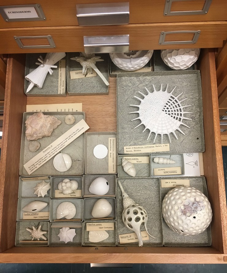
Václav Frič (1839-1916) was a natural history dealer based in Prague. He developed a series of 100 plaster of Paris models of Foraminifera (1861), as well as the stunning papier-maché models of Radiolaria (listed in his catalogue of 1878). He worked closely with Ernst Haeckel.

A selection of Frič’s models in the Museum’s stores

The Frič models oscillate between visible and invisible, illustration and model, art and science, philosophy and theory. They bear witness to a key period in the history of science when they were used to give tangible shape and proof to Charles Darwin’s poignant phrase:  “[…] from so simple a beginning endless forms most beautiful and most wonderful have been, and are being, evolved.”

Through the models we can “grasp” microorganisms that have been around for over 500 million years; organisms that truly have stood the tests of time.

Petri dish to puppetry

Spheres, spirals, rods, corkscrews… bacteria come in strange and beautiful shapes. Our Bacterial World exhibition (19 October 2018 – 28 May 2019) tells the untold story of life on a microscopic scale, and a recent Museum project brought together a research scientist, a group of school students and an artist to explore the patterns, textures and forms of beautiful bacteria. This science and art collaboration led to the creation of three fabulous bacteria-inspired puppets.

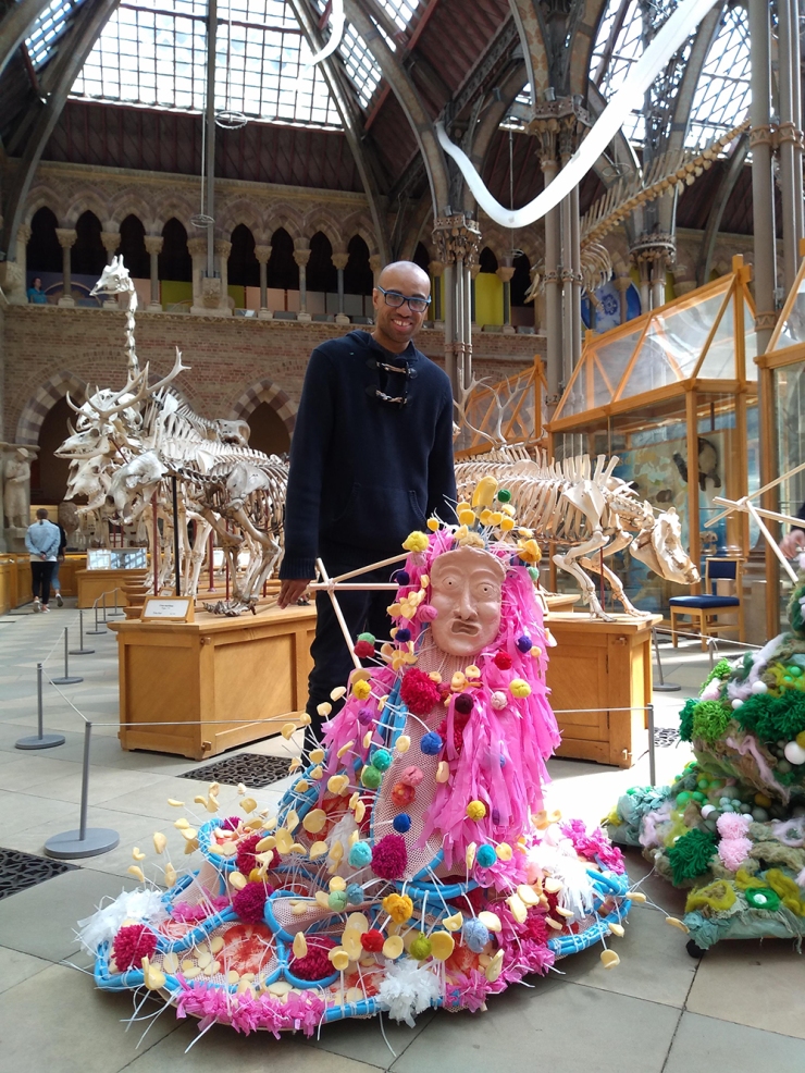
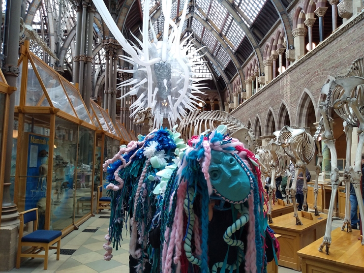
Volunteers and puppets in the museum
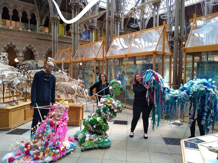
The puppets let loose in the Museum. Volunteers Tayo, Chantelle and Humaira (hidden behind the blue puppet!), with Carly from the Museum’s public engagement team.

Our Public Engagement team worked with Iffley Academy, a school for students with special educational needs and disabilities in Oxford. The pupils were from the brilliantly-named ‘Jackson Pollock’ class and they fully embraced the bacteria theme, through museum visits, workshops and classroom activities.

As well as visiting Bacterial World, the students had a workshop with Dr Frances Colles, a microbiology researcher from the University of Oxford, where they learnt about the importance of bacteria in their lives. As well as working with the students to create their own bacteria superheroes, Fran talked about her own work and took part in a Q&A, where the students made the most of quizzing a real, live scientist.

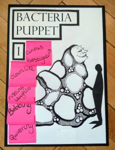
One of the character boards that Georgina created with the students

Next, the students spent two days with artist and puppet-maker Georgina Davy, who gave them the chance to experiment with a variety of textiles and techniques, including Japanese shibori dyeing, fringing, plaiting and knotting. The children even created latex faces to ‘personalise’ the bacteria. The pupils worked with Georgina to gather ideas and create mood boards and ‘characters’ for each puppet. She then used these individual pieces to build three giant, bacteria-inspired puppets.

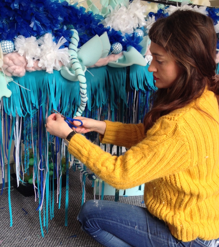
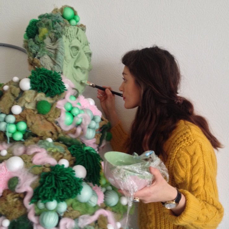
Georgina Davy in her studio, working on the bacteria puppets

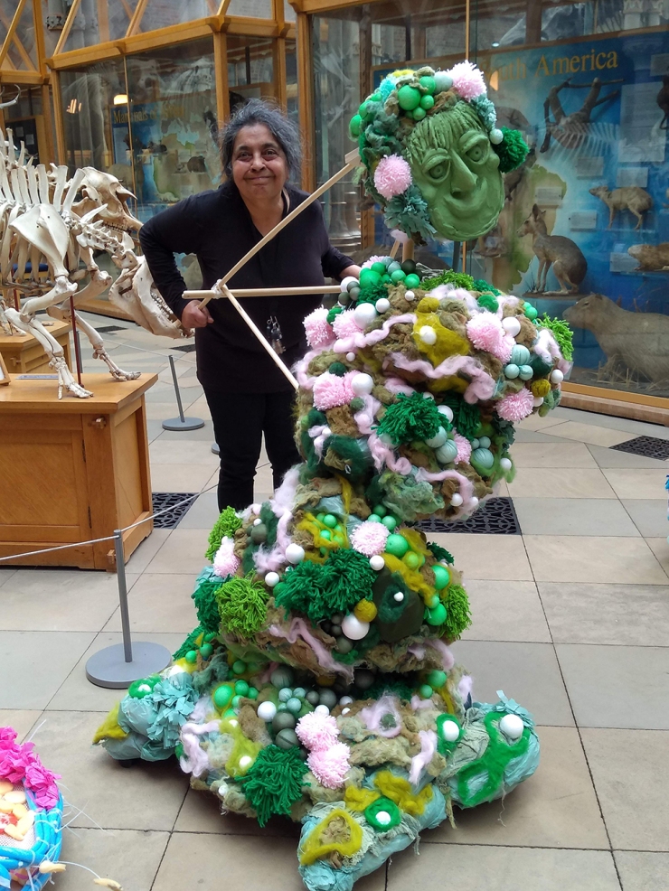
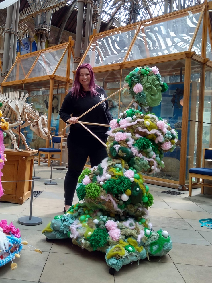
Just like the real bacteria that inspired them, the final puppets all have distinctive appearances and styles of movement. One is tall, green and plodding, another is pink, bobbing and quivering. The long, winding Chinese dragon-style puppet is slinky and searching. An artistic interpretation of bacteria, in motion.

Georgina Davy got a lot out of the collaboration and says:

This project has been the most unusual and marvellous project that a puppet maker could work on. Drawing upon scientific information from museum and academic staff that is enhanced and brought to life by students’ imaginations.

This project is unique in that the physical 3D puppet outcomes come from an almost entirely invisible world. Bacteria operate on an unfathomable microscopic scale. I am still finding it remarkable trying to envision this microscopic galaxy of bacteria taking place around us everyday in riots of colour, shape and movement. We cannot see the surreal bacteria forms that wriggle, bounce and swell around us, but they are there, some even tumbling around in forms like Chinese calligraphy. Their secret world is only unlocked by the microscope.

Once the puppets had been revealed to (and played with by) the students, they were transported to the Museum for the finale of the project – a public performance. On Saturday 11 May, three brilliant volunteers, Humaira, Tayo and Chantelle, showed off the work of Georgina Davy and the Jackson Pollock class to Museum visitors. The puppets twisted, shook and wiggled through the aisles, accompanied by percussion – drums and shakers courtesy of volunteers and visitors joining in with the performance.

If you’d like to see more about the Beautiful Bacteria project, we’ve put together a display in the Museum’s Community Case, where you can see original works by the Iffley Academy students. Until 6 August 2019.

The Beautiful Bacteria project was funded by BBSRC.
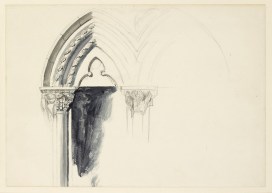
 

Ruskin 200 Art Competition

By Michelle Alcock, Front of House Deputy Manager

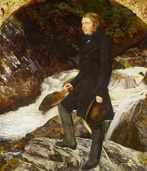
To celebrate the Museum of Natural History and the creativity it inspires, we have launched the Ruskin 200 Art Competition. It opened on Friday 8 February 2019 coinciding with the bicentenary of the birth of John Ruskin; an artist, social thinker, philanthropist and art critic of the 19th century. During the Victorian era, Ruskin’s views advocating for drawing from direct observation, both in his studies of Gothic architecture, and in his use of a detailed descriptive approach to depict nature in art, heavily influenced the design of the Museum.

WA2013.67 John Everett Millais, ‘John Ruskin’
Image © Ashmolean Museum, University of Oxford.

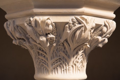
His encouragement led to artists, architects, craftsmen and scientists working together to design the Museum. As a result, they created the neo-Gothic building that stands today as a work of art and a vision of nature in its own right. The Museum’s architecture, decorative details, and collections have served as a source of inspiration for many since it opened in 1860.

Details in the Museum’s architecture, such as this carved capital, were inspired by nature and today provide further inspiration for visiting artists

This year marks the perfect opportunity to showcase the artwork of our visitors. Personally, working on the Front of House team here, I see what an inspiration the building is to our visitors. Every day we spot people of all ages setting up stools, with pencil and sketchbook at the ready, drawing in the Museum. There is so much potential inspiration; beetles carved in stone, vibrant birds’ feathers, glittering gemstones and the intricate decorative ironwork of the building, to name a few.

It is always exciting to see so many of our visitors engaging with the Museum in a creative way, but we rarely see the finished product. I’ve always wanted to know what artwork is created from this point of inspiration. Is it the starting point of a vibrant painting, an intricate pastel drawing or a graphic mixed media collage? The list of possibilities is endless.

A visitor captures the Allosaurus skull on one of our Sensing Evolution tables

Whatever your choice of creative expression, we want to see your interpretation of the Museum and what inspired you, whether it’s the architecture or the collections on display. If you are an amateur or professional artist, and over the age of sixteen, we would like you to submit your artwork to the Ruskin 200 Art Competition.

The competition is open for four months. Do send us images of your final artwork before the closing date of 19 May 2019. Selected artworks from each of the four entry categories will go on display in the Museum during the busy summer holidays.

A visitor taking part in creative activities during our special drawing weekend

Throughout 2019, we’re also running a programme of drawing activities to celebrate Ruskin’s bicentenary. It began with the Ruskin Drawing Weekend on 9 and 10 February, which included lots of different activities to begin the creative process. Look out for our Ready, Steady, Draw! workshops for younger artists coming in May too.

The full competition guidelines, along with further information on the Ruskin-related events we’re running this year, can be found on our website.

Top banner image: WA1931.47 John Ruskin, Design for a Window in the University Museum, Oxford. Image copyright Ashmolean Museum, University of Oxford


Writing from experience

The Museum’s building and collections provide inspiration for scientists and artists alike, often acting as a springboard for the creation of new work. Following a year here as one of three poets-in-residence, Kelley Swain returned to lead a session with Oxford Scholastica students, showing how museum objects can inspire creative writing.

by Kelley Swain

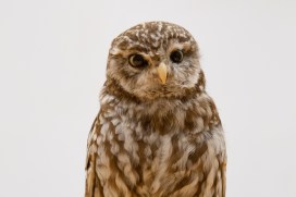
The experience of looking at the taxidermy Little Owl (Athene noctua) provided inspiration for Tallulah’s poem

Delving into the archives and behind-the-scenes stores, meeting researchers and conservators, and finding inspiration in the architecture, history, and collections were all part of my residency at the Museum during 2016. I’ve always written poetry inspired by the history of science and its fascinating objects, and I have come to appreciate museum objects not only as inspiration for my own poetry, but as teaching tools, or “object lessons” to inspire others.

It was lovely to be asked to lead a new series of these “object lessons” for a group of summer school students at Oxford Scholastica. Some of them had never encountered taxidermy, let alone a room full of articulated, stuffed, and preserved specimens. Awe abounded – both its wonder and, for some, its horror. It was a great opportunity to teach the students not only poetry, and why writing poetry inspired by museum objects can be moving, thoughtful, and important, but also to teach them about conservation and preservation.

Here we share the work of 13 year old Tallulah Xenopoulos, who created this poem following an encounter with a taxidermy owl during the workshop:

Stupid dead owl.
The wooden door opens slowly, and, although there’s a green stone with bumpy edges and
shiny sides, a jar filled with silky insects and a board with beautifully painted butterflies.
Both your eyes land on the owl.
His feathers brush down his back and he stares down at his lightly spotted blanket where his
delicate legs connect and hatch onto the bumpy branch.
His eyes
And his beak
And legs
And nails
He stares at you almost like he knows what you’re thinking – which is dumb because he’s
dead – but he scares you and fascinates you at the same time.
A piece of dust has fallen beneath his eye and I bet he’d love to just brush it away, cause
he’s like that.
But also.
He’s an owl.
A stupid.
Dead owl.
With nothing but stuffed insides and scrawny legs.
And a heart. A dead heart which they slipped out and replaced with stuff.
-”do you think they stuffed him alive?”
The boy next to you whispers. You don’t reply. But the thought of death. And of his feathers
falling the second he felt the blood rushing through him go cold and dusty, travels across
your mind.
“Do you think he knew he was about to be?” you answer
Because the poor clueless animal looks as if he knew nothing.
knows nothing.

Kelley Swain’s own poetry from the Museum residency is featured in Guests of Time, a beautiful hardback volume edited by Prof John Holmes which features new work by John Barnie and Steven Matthews, alongside 19th-century poetry from writers linked with the early days of the Museum. Together, the poems in this anthology are a tribute to the Pre-Raphaelite origins of the Museum and a rejuvenation of its artistic legacy.

Imagining lost worlds

Earlier this year University of Plymouth illustration student Rachel Simpson teamed up with our research fellow Jack Matthews to ‘bring the oldest multi-cellular organisms back to life’. Rachel tells us about the process of working with some of the most ancient fossil material and reveals the results of her illustrations and modelling.

Illustration by Rachel Simpson, created in collaboration with the Museum

In August 2018 I was lucky enough to travel to Newfoundland, Canada with Dr Jack Matthews to learn about and illustrate some of the extraordinary fossils found there. A highlight of the trip was going down onto the fossil surface – known as the MUN surface – to look at examples of organisms such as Beothukis, Charnia and Primocandelabrum, all of which date from the Ediacaran period, over 550 million years ago.

The MUN surface is the location of the fossils that I had worked on for my university project. I had spent the previous months sketching, drawing and bringing these organisms back to life from silicon casts, so it was amazing to be able to see the real specimens in situ and to sketch from the fossil surface.

Sketching directly from the fossils also provided a new challenge as I was unable to control factors such as the lighting, which is crucial to seeing the fossils clearly. Nonetheless, I learnt a lot about drawing on location.

Sketching at the fossil surface

While visiting Port Union I was able to use some of the old printing presses held by the Sir William F. Coaker Heritage Foundation to create work inspired by the fossils I had seen in the surrounding area. I love using printmaking in my own illustrative practice so it was a great experience to get to use these old presses (image at top of article).

We also had the chance to give a radio interview and talk to the Port Union community about the work that Jack and I had done, showing how science and art can work together.

On my last day in Port Union I was invited by a local potter to make some ceramic representations of the fossils I had been drawing there. I created models of Fractofusus and Aspidella, and discovered that re-imagining something in three dimensions is a very different process to recreating it as a drawing.

Rachel created ceramic representations of some of the Ediacaran organisms

For the final three days of the trip we relocated from Port Union to Trepassey to visit the Mistaken Point UNESCO World Heritage Site. Here, I saw the highly preserved Fractofusus specimens and made some more sketches. Using a small hand lens I was able to draw all the details that are invisible to the naked eye.

Using a hand lens allowed Rachel to pick out details in the Fractofusus fossil

Drawing on location in Canada provided a better idea of the organisms in relation to other surrounding organisms, something that is more obscure when working from museum specimens. This definitely informed my practice and meant that artwork created after the trip was more representative of the science.

When I returned to England, I created some new prints inspired by my time in Newfoundland, the fossils that I saw, and the printing process I was able to use in Port Union.

A set of prints made by Rachel based on her work in Newfoundland