A new year, a nice new display case. You may already be familiar with the Presenting… series that we’ve been running since March 2013; it started as a way to showcase treasures from the Museum’s collection during our closure year. Something changing and engaging to see as you passed through our darkened museum into the Pitt Rivers. Since re-opening early in 2014, we’ve celebrated significant natural history anniversaries, shared some of the staff’s favourite objects and put on joint displays with other departments in Oxford University. Now, for 2015, Presenting… is getting a make-over.
Bush cricket, family TettigoniidaeAmoret from Life collections installs a letter from Darwin to Hope
Today we’ve installed a brand new Presenting… display, in a posh new case. With humidity control and UV protection, this standard-leading unit gives us the opportunity to showcase some of the really special and fragile specimens from the collections. We’re launching tomorrow with a display of insects collected by none other than Charles Darwin.
As well as showing off some specimens collected by the great man in Australia and Tasmania, Darwin’s Insects will tell the story of his close friendship with Frederick William Hope (1797–1862), founder of the Hope Department of Entomology in this Museum. Hope was one of the most eminent entomologists of his time and when Darwin collected insects he often turned to Hope to help identify them.
Preparing specimens in the Life collections
Darwin’s journey on HMS Beagle began in 1831 and towards the end of the trip he travelled around parts of Australia and Tasmania observing and collecting many species, including the insects you can see on display. They’re displayed in pill boxes similar to the type Darwin would have used to collect the specimens originally, and you can see Darwin’s handwriting on the tiny labels.
Ant lion, family Myrmeleontidae
Alongside the pinned insects, you can see one of Darwin’s letters to Hope, sent in 1837. He mentions insects that he collected between January and April 1836, which include the specimens on display. He is asking for Hope’s assistance, because so many of these insects are unknown to science. Hope was always keen to help identify new species and in another correspondence, from 1834, he promised to give Darwin “all assistance in my power” with this task.
The insects and letter will be on display from tomorrow (10 January) until 8 March. Pop in and take a look!
Rachel Parle, Interpretation and Education Officer
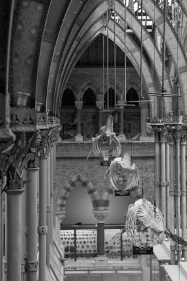
The sight of a huge Sperm Whale jaw soaring up to the roof is a familiar welcome to our visitors. But this spectacular specimen now has a companion. Resting against the opposite side of the cast iron column is a Humpback Whale skull.
The skull was donated back in the 19th Century by well-known scientist Professor Eschricht of Copenhagen.
Over the decades the specimen has been displayed in all sorts of places and positions around the Museum – laid flat on the floor, upright and on top of cases. Last year, as part of our Once in a Whale project, the specimen joined our other whale skeletons in undergoing some much-needed conservation treatment. You can find out the story of its big clean-up on the project blog.
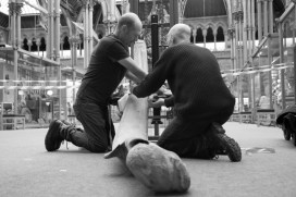
The skull is now displayed beautifully on a stand – but it was no mean feat to get it there. Bill Richey, the Museum’s Cabinet Maker, and Peter Johnson, Workshop & Maintenance, have carefully moved the specimen from the corner of the Museum where it was undergoing conservation treatment, reconstructed the complex structure and built a bespoke stand to support its huge weight. Here’s a step by step guide to rebuilding a Humpback Whale skull:
First, Bill used his years of experience in the Museum to build a display stand that perfectly held the complex contours of the bone. He scribed around the base of the skull, making a layer of MDF to fit each curve. Once he was confident of the perfect fit, he screwed them all together (see left photo), before returning to the workshop to square it all up, sand and paint the finished thing. He added a cushioning layer of Plastazote foam to the top surface, which would touch the skull.
Now to move the skull to its new location…
Because Bill and Pete had no idea how heavy the specimen would be, they decided not to take any risks and used the lifting machine to carry the weight. Keeping the specimen and themselves safe throughout the process was the most important thing.
Once they’d lowered the skull down to the floor, they used ratchets to hold it in place and secure the new base, using pieces of Plastazote foam to protect the sharp edges of the bone.
They then used the lift to tilt the skull into an upright position… to the point of no return. Bill says at this moment he was thinking;
I just hope it doesn’t crush Pete!
There was a sense of enormous relief at this point – the skull was upright, stable and fully supported by the new base. But Pete explained that the pressure was heightened throughout the process, because it was all so public. As it took several days, a lot of the work had to be done during normal opening hours, leading to a lot of intrigued visitors watching with great interest. No room to make a mistake without it being very obvious!
With the skull now safely in its new location, the construction began. Fitting the jaw bones was a serious jigsaw puzzle – working out which bits slotted in where and how to secure them safely to the column without any further damage.
To everyone’s relief, the Humpback Whale skull is now sitting proudly in its new stand, beautifully mirroring the neighbouring jaw. I’m sure Pete and Bill are hoping it won’t need moving again for quite some time…
Rachel Parle, Interpretation and Education Officer
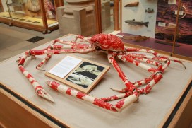
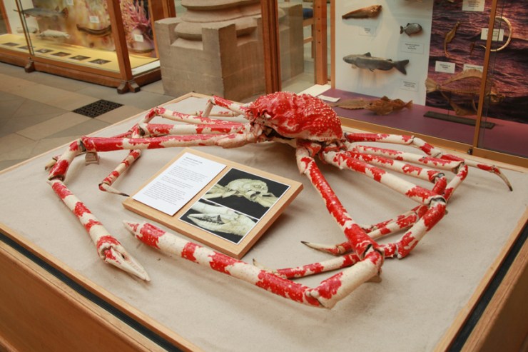
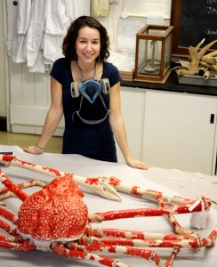
One of the most loved specimens in the Museum is the enormous Japanese Spider Crab. It’s been on display for over 100 years, so it’s unsurprisingly shown serious signs of deterioration. In July, staff in our Life collections decided that the crab should come off display and undergo conservation treatment. Bethany Palumbo, Conservator in Life Collections, took charge of this famous specimen.
The spider crab looking washed out after 100 years on display
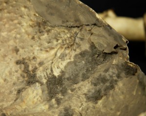
The most obvious damage was the loss of colour – the natural carotenoid pigments had completely faded due to decades of continuous light exposure under the glass roof. However, once it was taken into the laboratory for a closer look, Bethany soon realised that there were actually many areas that were fake, composed of old materials such as acidic cardboard, newspaper and even carved wood.
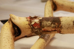
Some of the old filler material in a leg
These restoration efforts were causing more harm than good, deteriorating and damaging the natural shell material. The whole specimen was loosely held together with animal glue, PVA adhesive and, in some areas, tough wire which was cutting through the shell.
The first step for our conservator was to check the Museum database for information about the specimen, such as when it was donated and by whom. But unfortunately the specimen has no record, nor is it accessioned into the Museum’s collection. Although frustrating, this was important information. As it had no scientific data, Bethany could give this specimen more extensive conservation treatment, without compromising its scientific or historic integrity.
Bethany decided that treatment would consist of cleaning, the removal and replacement of old, deteriorating fill material and the restoration of colour to the shell, making the specimen true to life. These treatments, with the exception of the cleaning element, would be completely reversible.
Dirt build-up on the underside of the shell
Work began by taking the specimen apart to clean and treat each section. Sections were gently vacuumed and a moist cloth used to wipe away 100 years of embedded grime. Bethany removed old fill material, softening it with water vapour to allow it to be easily peeled away.
Newly filled and coloured leg
The next task was to create replacements for the missing sections. Bethany used a combination of acid-free tissue and closed-cell polythene foam. Intricate areas like the claws proved more challenging. Replacements were sculpted free-hand from Plasticine, moulded in silicone and finally cast in Jesomite composite plaster. They now look pretty close to the real thing and are a big improvement on the old wood and paper versions.
Replacement parts made from Jesomite plaster
Before it was ready to go out on display Bethany replaced the faded colour. Japanese Spider Crabs are bright red and white in life, but ours had become washed out beige.
Conservator Bethany with the finished Spider Crab
Photographs of Spider Crabs were used as a reference for the colours, and Bethany also spoke to crustacean experts in the Museum to make sure it was accurate. She used an airbrush and acrylic inks, selected for their high UV resistance. The shell was coated with a barrier layer to allow the ink to be removed in the future, if needed.
Airbrushing the specimen was the most time consuming element, as it required multiple layers and various brushing techniques to make the crab look true to life. Once completed, Bethany gave the crab a final protective coating, providing good water resistance, ready for the next time it needs a good clean!
Rachel Parle, Interpretation and Education Officer
One of the most uplifting projects here over the past year or so (literally, as you’ll see) has been the conservation work on the five whale skeletons suspended in the court. The skeletons are beautiful, the process was intricate, and the whole thing was rigorously documented on our accompanying Once in a Whale blog.
The work inspired filmmaker Robert Rapoport to record some eerily captivating footage of our conservators at work, and the project itself was Highly Commended in the Museums + Heritage Awards.
At completion, the whales were raised once again into the vaulted space, but this time rearranged in size order and staggered in their distance from the ground. Each has its own spotlight, creating an impressive display, especially once darkness falls outside.
But there was a final element to the displays that has just been installed: information panels containing details about each of the species suspended above, along with drawings and paintings created for us by artists Nicola Fielding and Claire Duffy.
Claire’s paintings of the whales have been used in a scaled schematic of the display, each ‘fleshed out’ to give an impression of the whale in its full form; and Nicola’s accurate recreations of the skeletons are featured in a second panel which gives details of the conservation project itself.
A schematic drawing of the whales suspended in the court, along with further information about each species
Nicola is something of an old hand when it comes to making drawings for the Museum – her work is featured on lots of our family trails already. But the whale project seems to hold a special place in her heart:
I could write a short essay about how much being involved in the whale project meant to me. I’ve always been mesmerised by cetaceans and by the mythical status they can have. In a museum, hanging alongside dinosaur skeletons, they can seem like something we only know from pictures and imaginings. But cetaceans are of course still living, breathing and can be found in all corners of the worlds oceans. Even around the UK there are so many species to be found.
So I was really excited to be involved in a project that would allow the Museum to make the most of its incredible skeletons, and to make sure all the knowledge we do have about them is shared.
One of the panels in the whale aisle gives details of the conservation project
We hope the new information panels at each end of the whale aisle will encourage visitors to look up and perhaps share in Nicola’s wonder for these amazing creatures, many of which were almost hunted to extinction during the periods of intense industrial whaling.
Finally, if if you like the look of these paintings, there’s a last chance to see some of Claire Duffy’s other work in her Avifauna show at the Old Fire Station in Oxford, which runs until Saturday 8 November.
The ecological importance of bumblebees has become more widely appreciated in recent years, thanks to environmental campaigners and reports of species decline, and even some extinctions, in the UK.
To look at this issue, we have recently teamed up again with arts-science organisation Pale Blue Dot, which is launching a new research project to investigate why some species of bumblebee are declining and to raise awareness about the ultimate impact this has on people.
Here, Pale Blue Dot co-founder Jane King explains how the Bees & Weeds project brings together art students, public engagement, the Museum’s collections and a leading bumblebee scientist…
*
On 9 September we launched our latest project – Bees & Weeds – with the Museum of Natural History, building on our previous collaboration for the Lost & Found exhibition. We were joined by over 50 art students from Banbury & Bicester College to highlight the plight of the bumblebee, revealing how its decline is impacting everything from what we eat to where we live and work.
Amoret Spooner displays drawers from the collections
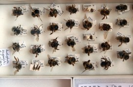
The students spent some time looking at methods of insect labelling and notation, before heading behind the scenes with entomologist Amoret Spooner to the Huxley Room, the location of the Great Debate on Darwin’s Theory of Evolution, which took place in 1860.
Amoret provided an insight into taxonomy – the science of species classification – as well as her work on the conservation of specimens. We visited the huge archive of bee specimens and learnt about some of the research that scientists are currently carrying out on UK bumblebee species to help prevent further decline.
Student sketches of labelling and notation
During spring 2015, the art students from the Banbury & Bicester College, as well as students from Oxford Brookes University, will make and install hundreds of cycle seat covers on bikes in and around the Oxford city. The seat covers will carry messages about bumblebee decline in the UK countryside, showing how much we depend on their pollination services, which far outstrip those of the honeybee in their value to UK food production.
We are also working with Professor Dave Goulson from the University of Sussex, one of the world’s most important bumblebee scientists. He will present his research showing how well bumblebees are doing in gardens compared to the countryside, as well as the optimum range of flowering plants needed to help them thrive. Dave’s book, A Sting in the Tale, is already a best-seller, and the sequel – A Buzz in the Meadow – was published on 4 September.
Dave will also be speaking about his new book at the Museum on Thursday 9 October at 7pm. Book your tickets for that via Waterstones here.
Artwork from the Bees & Weeds project, together with cycle seat covers and bike paraphernalia, will be on show and for sale in the Old Fire Station in Oxford from next spring. If you cycle in Oxford, you may be lucky enough to receive one!
Pale Blue Dot is an arts-science organisation helping scientists to communicate their research to the public. It promotes an interdisciplinary approach to learning, living and working through exhibitions, publications and happenings.
They say a picture is worth a thousand words, and at the Museum we make thousands of pictures: pictures to document, pictures to investigate, and pictures to wow. We use a lot of different imaging techniques too, from standard close-up photography to scanning electron microscopy, which reveals the most minute details.
To coincide with the final week of the Wildlife Photographer of the Year exhibition here, on Saturday 20 September we held a new adult workshop to give people some hands-on experience of some of these processes. Imaging Techniques in Modern Natural History gave participants the chance to get up close to some wonderful specimens and make their own images to take home.
I had planned to review the day here, but Rose Parkin, who took part in the workshops, very helpfully sent in her own write-up of the sessions. So here’s a special guest post from Rose, along with some pictures taken by people on the day.
*
By Rose Parkin
When I signed up for the digital imaging course I expected a fairly dry, tech-heavy day. Instead, the experience was really exciting. Not only did it provide hands-on experience of viewing and recording images with new technology, it also gave me a brief glimpse behind the scenes of my favourite museum.
Laser Scanning and Digital Modelling For our first session our small group was led through a maze of corridors by Sarah Joomun, the Documentation Officer, to the laser scanning lab. It sounded a bit futuristic, and it turned out that it looks that way too. Sarah popped a fossil onto a mount, clicked a few buttons and red lasers appeared, scanning the fossil’s surface while it rotated. After ten minutes the first 3D image of the fossil was produced – a beautiful net of triangles, which looked like a teleporting object in a science fiction film.
Laser scanning Image copyright Tom Nicholson-Lailey
Sarah turned the fossil and scanned it again. The challenge was then to fit these two images together to make a complete 3D model. Amazingly, this technique enables other palaeontologists around the world to see and replicate, with the use of a 3D printer, the exact size and shape of a fossil without it ever leaving the museum.
Multi-plane Microscope Photography Our next session was upstairs, with artist-in-residence and photographer Katherine Child. Even though we were close to the main corridor of the museum it felt like a real working space, crammed full of equipment and insect specimens. Katherine had chosen the tiniest of insects for us to photograph with the multi-plane microscope. It looked like a small seed with some barely visible limb-like protrusions.
Multiplane photography. Image copyright Rose Parkin
But under the microscope a wonderfully strange insect became visible, with the most bizarre appendages and bright orange legs. While the microscope already showed a great deal of detail the multi-plane photography captured an incredibly crisp image. The microscope takes large numbers of photos of the specimen, using different focal planes each time, then the focussed elements are all stacked together to produce a crystal clear photograph.
Once we’d chosen and photographed some other insects from the collection and poked around the room a bit (finding a disturbing collection of large pickled spiders), we were taken on a tour of the entomology department. Katherine led us through corridors of offices and labs, up to a stunning store room that felt almost church-like, with rows and rows of cabinets full of fascinating insects.
Scanning Electron Microscopy After lunch we had a laboratory session with museum director Paul Smith to look at sand under an electron microscope. Luckily, that was much more exciting than it sounds! The sand was taken from Dog’s Bay on the west coast of Ireland and is rich with a wide range of tiny fossilized organisms. Paul showed us how to carefully select individual microfossils from a tray using just a microscope and a paint brush.
Professor Paul Smith demonstrates the scanning electron microscope. Image copyright Rose Parkin
We then viewed some of the microfossils using a scanning electron microscope. This allowed us to see an incredible level of detail. The microscope was so powerful that we could see hair holes in a fossil the size of a grain of sand.
DSLR Macrophotography My final session was a crash course in macrophotography. Held in the seminar room, the low lighting and floor-to-ceiling collection of specimens lent an almost eerie feeling to the set-up.
Macrophotography. Image copyright Keith BarnesBearded dragon. Image copyright: Rose Parkin
Once prepped, we were let loose on four separate camera setups. Being able to choose and shoot at our own pace made this feel like a really creative experience. The help given by professional photographer Keith Barnes and public engagement officer Scott Billings was perfect – very hands on but not patronizing (despite my lack of DSLR experience).
With this digital imaging course the museum has created a really exciting snapshot of the work that goes on behind the scenes, reinforcing the fact that this impressive place is much more than just an ordinary museum.


 The skull was donated back in the 19th Century by well-known scientist Professor Eschricht of Copenhagen.
 First, Bill used his years of experience in the Museum to build a display stand that perfectly held the complex contours of the bone. He scribed around the base of the skull, making a layer of MDF to fit each curve. Once he was confident of the perfect fit, he screwed them all together (see left photo), before returning to the workshop to square it all up, sand and paint the finished thing. He added a cushioning layer of Plastazote foam to the top surface, which would touch the skull.