How we got bigger, more vulnerable brains

This article is taken from European research magazine Horizon as part of our partnership to share natural environment science stories with readers of More than a Dodo. For more on the development of the brain see our Brain Diaries exhibition site.

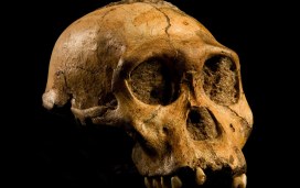
One of the major features that distinguishes humans from other primates is the size of our brains, which underwent rapid evolution from about two to three million years ago in a group of our ancestors in Africa called the Australopithecines. During this period, the human brain grew almost three-fold to reach its current size. Scientists know this from skull remains, but have puzzled over how it happened…

This year, the mystery was partially solved by Professor Pierre Vanderhaeghen at the Flanders Institute for Biotechnology in Belgium. Prof. Vanderhaeghen, who was conducting his work as part of the GENDEVOCORTEX project, went on a hunt for the genes that drove the growth of human brains.

Scientists had suspected that brain expansion began in our human ancestors when they evolved genes that are switched on in the foetus, when a lot of key brain development occurs. Prof. Vanderhaeghen therefore looked for genes present in human foetal tissue, but missing from our closest living relatives, apes.

His lab discovered 35 hominid – present only in apes and humans – genes that were active in foetal brain tissue. They then became intrigued by three specific genes – all similar to NOTCH genes, an ancient gene family involved in sending messages between cells and that are present in all animals. They found that the three new genes, collectively named NOTCH 2NL, were created by a “copy and paste error” of an original NOTCH gene.

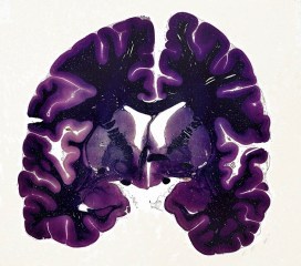
This error created entirely new proteins which likely helped our ancestors’ cerebral cortex to balloon. This is the part of our brain responsible for our language, imagination and problem-solving abilities. Scientists at the University of California, Santa Cruz, have also identified the NOTCH 2NL genes in DNA from Homo sapiens’ extinct cousins – the Neanderthals and Denisovans.

(The NOTCH 2NL) genes are only present in humans today. They were also present in Neanderthal DNA, but not in chimpanzees
Prof. Vanderhaeghen

Evolution
These genes control the growth rate and differentiation of brain stem cells – the starter cells that multiply and give rise to all neurons in our brain – causing them to seed more nerve cells, which in turn helped to expand brain size. The genes likely led to more neurons and brain tissue in our ancestor’s descendants – including Neanderthals, Denisovans, and modern humans.

Prof. Vanderhaeghen’s research could also help to provide new insights into brain disorders. The US researchers linked genetic faults in DNA that were very similar to NOTCH 2NL, to children born with enlarged brains or small brains. Many of the new human-specific genes are located in a small area of our genome that plays an important role in brain size, according to Prof. Vanderhaeghen.

As DNA in this area closely resembles another part of the genome where it was originally cut and pasted from millions of years ago, errors are more likely, said Prof. Vanderhaeghen. “Patients who have (inherited) deletions in this area tend to be at risk of developing schizophrenia, whereas patients with duplications are more at risk of autistic spectrum disorder,” he said.

Prof. Vanderhaeghen is now studying some 20 of the remaining human-only genes to see how they contributed to the evolution of the human brain.

Something like 40-50% of the Neanderthal genome can still be found in people today.
Prof. Svante Pääbo, Max Planck Institute for Evolutionary Anthropology, Leipzig, Germany

The use of genetics to study human evolution in this way is helping to transform our understanding of how our own species compared to our ancestors. Traditionally, scientists have studied extinct species by looking at the fossilised remains of their bones. This was how they discovered the existence of Neanderthals, the extinct human species that lived across Europe and much of Asia before vanishing around 40,000 years ago.

In the last decade, however, scientists have begun to look at the DNA inside these bones. Professor Svante Pääbo, director of the Max Planck Institute for Evolutionary Anthropology in Leipzig, Germany, has led the way in sequencing DNA of these extinct humans from small bone fragments.

This allows scientists to compare modern human DNA with that of extinct humans, rather than just living relatives like chimps. Already, the work has revealed some surprising findings – our own species appears to have interbred with some of these ancient relatives during our history.

Ancient humans
Scientists have found that the DNA of every person outside Africa is 1-2% Neanderthal, meaning that these extinct human relatives had offspring with our own ancestors.

An international consortium of researchers is sequencing the 3 billion bases that make up the genome of our closest relative – the Neanderthal. The sequence is generated from DNA extracted from three Croatian Neanderthal fossils using novel methods developed for this project. Image credit – Frank Vinken for Max Planck Society

“Different people tend to carry different pieces of the Neanderthal genome,” said Prof. Pääbo, who is undertaking a project called 100 Archaic Genomes to decipher the DNA of ancient human individuals. “Something like 40-50% of the Neanderthal genome can still be found in people today,” he said.

According to Prof. Pääbo, we retained some of this DNA because it offered an advantage to our ancestors. “Some (of this retained DNA) has to do with the immune system, presumably helping us to fight off infectious diseases.”

The power of genetics to unravel the history of human evolution took a new twist in 2010 after Prof. Pääbo’s lab sequenced DNA from a finger bone fragment found by a Russian archaeological team in a remote Siberian cave.

The analysis revealed the bone belonged to a previously unknown human relative, now called Denisovans after Denisova Cave where the bone was found. This mysterious ancient human species lived at around the same time as Neanderthals, but further east into Asia.

Last year, Prof. Pääbo’s group published DNA sequences from a tooth found in the cave – the fourth ever Denisovan discovered. We now know Denisovan DNA carries more variation than Neanderthal DNA, leading scientists to conclude that they were more widespread than the better-known Neanderthals.

Denisovans left a more impressive stamp on some of us than Neanderthals, according to Prof Pääbo. Their DNA can be found in people across Asia today, while indigenous peoples of Papua New Guinea and Australia may carry up to 5%. Tibetans also carry some Denisovan DNA in their genomes, which has helped them adapt to life at high altitudes where there is little oxygen in the atmosphere.

Prof. Pääbo and his colleagues will soon publish their third high-quality genome – where almost the entire DNA sequence is intact – of a Neanderthal from Siberia. A deciphered genome of this quality allows for better DNA comparisons and could tell us more about the evolution of important genes – such as those linked to the development and function of the brain. It will add yet another puzzle piece to help us understand the history of our closest extinct relatives, according to Prof. Pääbo.

“There may even be other forms of extinct humans out there to be discovered by studying the DNA of the (ancient) bones we find,” he said.

Top image: The skull of a Australopithecus sediba, a species of Australopithecines, who were our ancestors and whose brains started to grow two to three million years ago. Image credit – Australopithecus sediba by Brett Eloff, courtesy Profberger and Wits University is licensed under CC BY-SA 4.0.

*

This post Genetic error led humans to evolve bigger, but more vulnerable, brains was originally published on Horizon: the EU Research & Innovation magazine | European Commission.

The winning brainwave

If you could create an experiment to learn more about the human brain, what would you investigate? We posed this question in our Big Brain Competition last year, as part of the Brain Diaries exhibition with Oxford Neuroscience, and received a whopping 800 entries!

For the competition, Oxford University neuroscientists offered people the chance to use the state-of-the-art MRI scanner at Wellcome Centre For Integrative Neuroimaging at the John Radcliffe Hospital to investigate a burning question about the brain. We had ideas from the young and old, and by visitors from all around the world suggesting brilliant questions and some fascinating experiments.

Memory_fMRI
Functional MRI image of the human brain using the MRI Scanner

To judge all the ideas, entries were split into categories: feasible experiments, unfeasible experiments, under 18s, and questions about the brain. WIN researchers compiled a long-list for each, which was ranked by a panel of neuroscientists and people from the museum to reach the eventual winners.

Sadly, only one experiment could be carried out, so an overall winner was picked from the ‘feasible experiments’ category. The winning experiment was suggested by Richard Harrow, who wanted to understand how the brain identifies voices.

A person is put in the MRI scanner with headphones on.  They are shown a photo of a person familiar to them, either a friend, family member or celebrity.  Then, in their headphones they are played the voice of a person, but the voice is either sped up or slowed down.
They are required to say whether the face on the photo matches the voice they have heard. What happens in the brain when this confusion of audio and visual information is occurring? Will the brain find a way to identify the vocal signature of the voice, even if distorted, and be able to say with conviction if the photo and the voice are a match?
– Richard Harrow, winning competition idea

IMG_4498 (2)
Competition winner Richard Harrow was interviewed alongside neuroscientist Dr Stuart Clare during the live streaming of the experiment

On the day of the experiment, the winners and runners-up headed over to the WIN Centre to watch Richard’s winning experiment being conducted. The experiment was streamed live by Oxford Sparks and we had a clear result from the test, as neuroscientist Dr Holly Bridge explains:

The scans show that when you’re getting information that corresponds in both your auditory and your visual system you get a boost in your brain activity. We can detect that the brain does respond differently depending on whether or not you can match the face with the voice; it clearly has a lot to do with expectation.

Stuart and Holly
The brains behind the Big Brain Competition, Dr Holly Bridge and Dr Stuart Clare explained the results of the experiment on a Facebook Live stream

The scientists also wanted to answer as many of the other great brain questions as possible. So a series of articles picks out some of the broad themes in the competition ideas, including lifestyle, muscle memory and stress. Researchers also answered more big questions live on Facebook during this year’s Brain Awareness Week.

We sorted the many entries in the Big Brain Competition into themes such as vision, lifestyle, and language

Thank you to everyone who suggested an experiment or asked a question; it made for a fascinating conclusion to the Brain Diaries exhibition, and has definitely increased the amount of brain activity from staff across the Museum and Oxford Neuroscience… if only there was an MRI scanner for us to see it!

IMG_3989
Winners of the Big Brain Competition at the Museum of Natural History with the neuroscientists. Left to right: James, Holly, Stuart, Misha, Richard, Lily, and Heidi

You can still get involved with the Big Brain Competition by trying the winning experiment at home.


The crucial cortex

Lance Millar_Developmental Anatomy_14Oct2017.jpg-large

University of Oxford PhD student Lance Millar recently ran one of our Brain Spotlight events as part of the Brain Diaries exhibition programme. Here, Lance explains his research into neurodevelopmental disorders and possible treatments.

The brain has always been a fascinating organ for me. It is the site of our intelligence, our problem-solving and social skills, and it allows us to connect our senses to the world around us.

The large, folded outer part of the human brain is called the cortex, and is responsible for decision-making, language, face recognition, and a lot of the other things that I like to think are what make us human. The word cortex comes from the Greek for husk or outer shell, which underestimates the importance of what the cortex does.

Humans can survive with damage to the cortex, but depending on the part of the cortex that is damaged, a range of disabilities can result. People who have had a stroke can lose part of their cortex, leading to limb paralysis, loss of speech, or loss of memory, depending on the site of the damage.

B0009564 Human brain, coronal section, LM
Cerebral cortex – Professor Michael R Peres – Wellcome Images

Some people are also born with a developmental problem in the cortex, and are said to have a neurodevelopmental disorder. Such conditions are thought to include autism, schizophrenia, ADHD, and even dyslexia – all fairly common conditions. The damage to the cortex is subtle and complex in these conditions, and scientists are still working out exactly what happens to the brain during its prenatal development.

I am studying one particular neurodevelopmental disorder caused by lack of oxygen at birth. It is known to medical specialists as neonatal hypoxia ischaemia. The image on the right shows a cross-section MRI scan of a normal newborn human brain, alongside some babies who have been damaged by oxygen deprivation. You can see that the brains are smaller, the cortex is less folded and it takes up less space inside the skull.

Woodward
MRI scans of normal newborn brains alongside those of babies who have been damaged by oxygen deprivation. Image: Woodward et al., New England Journal of Medicine, 2006

Scientists still don’t know how to protect the newborn brain from these injuries. Some are caused by inflammation which is a normal response to illness, but can wreak havoc in the confined space of the skull. Some is caused by the presence of free radicals, which are thought to contribute to ageing and organ failure, as the newborn brain doesn’t have many antioxidants to fight these chemicals. It’s also possible that the electrical signals that neurons within the brain send to each other contribute to the damage when there isn’t enough oxygen to feed them.

So what can we do to treat oxygen deprivation at birth? One breakthrough treatment currently available is known known as hypothermia. In this technique, the baby is cooled to 33℃ which slows down the brain-damaging chemical reactions which in turn protects the brain. This is currently the only treatment available, but I am involved in the study of possible alternatives.

We don’t want to introduce any drugs to the baby’s system as they might be harmful to normal development. So scientists are currently working on treatments which help the baby’s natural body proteins to protect the brain. I do this by looking at neurons under the microscope, and identifying proteins expressed by these neurons using fluorescent probes known as antibodies.

Neurons under the microscope
An example of neurons under the microscope. Image: Lancelot Millar

These neurons are expressing neuroserpin, a natural brain protein which decreases inflammation and cell death. I’m looking at exactly where neuroserpin is expressed in the brain, how it can be upregulated in response to oxygen deprivation, and how its chemical reactions could be used to protect the brain.

Another way to help people with neurodevelopmental disorders is to better understand how the cortex connects to other parts of the brain and how it can carry out complicated decisions. There is still so much to understand about the complexity of the human brain, and what seems like fundamental research could generate the springboard for new ideas for neurodevelopmental disorder treatments.

To explore the structure of the human brain and compare it to that of other animals see the Brain Diaries Brain Explorer below.


All about Alzheimer’s

University of Toronto research fellow Jacqueline Zimmermann recently ran one of our Brain Spotlight events as part of the Brain Diaries exhibition programme. To mark World Alzheimer’s Day today, here Jacqueline tells us about the neurophysiology of Alzheimer’s disease and the risk factors we can actively reduce to lead happier, healthier, and longer lives.

Almost all of us have in some way been affected by Alzheimer’s disease, which makes the quest for a cure that much more personal. An estimated one in nine people over the age of 65 will develop the disease, and this risk also increases with age, according to the World Alzheimer’s Report in 2015.

Brain Spotlight
Jacqueline Zimmerman’s Brain Spotlight on Saturday 16 September as part of the Brain Diaries exhibition series of events.

Due to chemical toxins, and increased longevity, the incidence for Alzheimer’s disease is on the rise. But the good news is that there is a lot that you can do to reduce your risk. At the John Radcliffe Hospital in Oxford, hundreds of scientists are currently working towards identifying the cause and the solution to the disease.

At the Brain Spotlight event at the Museum I presented images of ageing brains, and explained how brains affected by Alzheimer’s have reduced volume in the temporal lobe and the hippocampus, regions critical for language and memory respectively. Diseased brains will also often show reduced frontal lobe volume, which may reflect the changes in personality and the ability to engage in planning which area associated with Alzheimer’s. The overall volume of the brain is also reduced in sufferers because cellular changes lead to the death of neurons.

Brain Atrophy in Alzheimer’s disease
Brain Atrophy in Alzheimer’s disease. Note: overall brain volume is reduced, hippocampal regions and frontal regions are particularly affected, and ventricles are enlarged. Image: http://www.lookfordiagnosis.com

Recently, a number of genes have been identified that are related to early onset Alzheimer’s, which is quite rare and much more hereditary than late-onset Alzheimer’s. At the Nuffield Department of Clinical Neurosciences, where I am a visiting researcher, we are investigating a late-onset Alzheimer’s risk gene called Apolipoprotein 4 (APOE4), looking at how it relates to subtle cognitive impairments in middle-aged people. Working with the Oxford Biobank we are trying to determine which cognitive assessments may be most effective in predicting these impairments.

Jacqueline Zimmerman
Functional brain imaging using electroencephalography at Rotman Research Institute in Toronto. Image: Rotman Research Institute

Although some of us may be more susceptible to Alzheimer’s than others, there are a number of environmental factors that contribute, including air pollution or additives in our food, like nitrogen-based chemicals which are used to preserve and flavour processed foods. It is important to reduce cholesterol in the diet, eat plenty of fruits and leafy greens, and engage in frequent physical and mental exercise.

Though there is speculation about the effectiveness of ‘brain games’ and how they translate into improvements in cognition in the real world, there are certainly large benefits of keeping your brain active.


The Big Brain Competition

What happens in your brain when you receive compliments? And what’s going on in your mind when you watch your football team win a match? Does the brain respond differently when recalling music, compared to listening to it? All these questions, and more, have been posed in our Big Brain Competition

Coinciding with the Museum’s Brain Diaries exhibition, the Wellcome Centre for Integrative Neuroimaging is inviting you to ask your own question about the brain to be in with a chance to have it tested by neuroscientists using Oxford’s state-of-the art Magnetic Resonance Imaging (MRI) scanner.

The advanced MRI scanner at the John Radcliffe Hospital in Oxford is one of the strongest in the world. It allows scientists to carry out functional MRI (fMRI) scans to see the brain in action. This mind-blowing procedure can reveal how the brain changes when learning a new skill or how it compensates when someone recovers from brain damage. It can also reveal which areas are used when people speak, move or laugh, to give just a few examples.

This fMRI scan shows how blood flows to the visual cortex region at the back of the brain when viewing a visually-stimulating checkerboard pattern
layouttile
Dr Stuart Clare of the Nuffield Department of Clinical Neurosciences is asking you for questions about the brain

Functional MRI shows when a brain area is more active by detecting the changes in blood oxygen levels and blood flow that happen in response to neural activity. The technique can be used to produce activation maps showing which parts of the brain are involved in a particular mental process.

The scientist behind the Big Brain Competition is Dr Stuart Clare, whose research involves pushing the technological boundaries of the fMRI technique to reveal new insights about how the brain functions normally and how it is affected by disease. There is still so much that the fMRI scans can bring to light, so Stuart is asking you for ideas!

Over several years of inviting people in to see the beautiful pictures that our MRI scanner can produce, I’ve been fascinated by the questions they have about the brain and whether you can see this thing or that thing in our fMRI scans.  With this competition we want to give people the unique access to our scanner and the chance to try an idea out for themselves.

When coming up with an idea for investigation there are a few practical things to bear in mind. Any activity has to be something people can do when lying down in the scanner and it has to be clear when they start and stop doing the activity. But Stuart is very open to ideas for experiments that they haven’t come across before – something that scientists really don’t already know the answer to.

The animation below explains how fMRI works and what it can do. So take a look, think up an experiment of your own and enter your idea via this form. The best one will be put into action by the research team and you will be able to watch the scans take place at the John Radcliffe Hospital yourself!

Credit: Mike Peckett

Great minds don’t think alike

Credit: Mike Peckett

Museums are a place for many things: inspiration, learning, conservation… the list goes on. But we believe that they should also be a forum for debate and discussion. One of our aims, as part of our Contemporary Science and Society exhibition and event series, is to bring controversial or challenging ideas to our visitors and to encourage a lively, informed and balanced debate.

Thomas Henry Huxley

Controversy is nothing new to this institution; there is a history of debate going right back to 30 June 1860, the year the Museum was founded. The Great Debate is believed to have been the first ever public debate on Charles Darwin’s theory of evolution – certainly the thorny issue of the time.

The debate is now notorious for the clash of ideologies between Samuel Wilberforce, the Bishop of Oxford, and Thomas Henry Huxley, a young biologist known as ‘Darwin’s bulldog’.

Samuel Wilberforce

Reports from the time are a little sketchy, but tempers are believed to have flared and insults were traded, climaxing with the shocking moment where Wilberforce compared Huxley’s grandparents to an ape. This was obviously outrageous to the delicate Victorian temperament, and people were believed to have fainted with shock!

This short video reveals how we think the debate may have gone (with a little artistic license thrown in):

The anniversary of the Great Debate falls next week, and this year it’s an extra special one. We’re reigniting the tradition of a good lively discussion with The Great Debate: Smart Drugs – Is It Cheating? On Thursday 29 June, Claire Fox of the Institute of Ideas and BBC Radio 4 chairs a multidisciplinary panel as in a debate about the ethics, fairness, and effects of so-called smart drugs and their impact on society.

Smart drugs is a name given to prescription drugs, typically used to treat disorders such as narcolepsy and attention deficit hyperactivity disorder (ADHD), which are now also commonly being used to improve cognitive ability and concentration. Some studies suggest that these drugs are now widely being used by university students, in a climate of increased academic and financial pressure.

Many students are said to see these drugs as a normal aid to study, but some experts have serious concerns about increasing levels of self-prescription and the long-term safety of their use, as well as the impact on competitiveness. Increased use in other areas of society may also have implications.

The panel for this debate includes world experts in the fields of neuroethics, evolutionary psychology, and philosophy, each representing different sides of this challenging subject.

Tickets are free, but you need to book your place. We can promise controversial opinions, expert insights and an eye-opening evening, but unlike Wilberforce and Huxley, we can’t necessarily guarantee that the panelists will be flinging insults about each other’s grandparents.