Some of the very oldest complex, macroscopic communities on Earth appear in the fossil record about 570 million years ago and record the presence of a group of organisms – the rangeomorphs – with an unfamiliar body plan that, at their ultimate extinction, was lost from life’s repertoire.
Rangeomorphs are characterised by a strange frondose branching anatomy, where large primary branches host smaller branches which themselves host smaller branches again. This arrangement appears to maximise the surface-area to volume ratio of the organism, rather like a lung or a gill would today.
The smallest known rangeomorphs are less than a centimetre in length, but they grew huge and the largest records indicate they could stand more than two metres tall. There is no evidence to suggest that rangeomorphs were able to move around, rather, they lived stuck to the sea floor in the deep ocean, far below the reach of light.
Despite this strange set of characters, there is growing consensus that rangeomorphs likely represent very ancient records of animal life. However, they lived at such a remote time in Earth’s history that they do not possess any direct living descendants. Given all this, it may not be a surprise to hear that we know relatively little about how these organisms made their living and came to dominate the ancient seafloors.
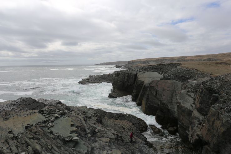
The UNESCO world heritage site Mistaken Point in Newfoundland, Canada, is one of the sites on which we find exceptionally preserved rangeomorph fossils. Photo: Alex Liu.
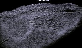
In order to better understand them, my co-author Alex Liu and I travelled to Newfoundland, Canada to explore the rocks which host these remarkable fossils and over the past few years we have made an unexpected discovery. We found that fine filamentous threads connect rangeomorph fronds of the same species, in some cases over many meters, though they are typically between two and 40 centimetres long.
An undescribed rangeomorph fossil with filamentous connections at the base of the frond. We find that this species of rangeomorph can be connected to each other over meters! Photo: Alex Liu.
It is possible that these filaments were involved in clonal reproduction, like strawberry plants today, but they may have had additional functions such as sharing nutrients or providing stability in strong ocean currents.
The discovery of the filaments means that we have to reconsider how we define an individual rangeomorph, and may help us understand how rangeomorphs (seemingly) rapidly colonised deep-sea environments. Either way, some reassessment of the palaeobiology of these unique organisms is certainly required!
Our current First Animals exhibition is extending its run until 1 September, and to mark the extension our Research Fellow Imran Rahman takes a look at how animal life in the ancient oceans was brought to life in our Cambrian Diver interactive installation.
One of the biggest challenges in developing the First Animals exhibition lay in visualising rare fossil specimens as ‘living’ organisms, transforming them from two-dimensional imprints in the rock into three-dimensional animated computer models.
Many of the specimens on display in First Animals were collected from sites of exceptionally well-preserved fossils called Lagerstätten. These deposits preserve the remains of soft-bodied organisms that are almost never seen in the fossil record; things such as comb jellies and worms, as well as soft tissues such as eyes, gills and muscles. Even so, most of these fossils are flattened and two-dimensional, which makes it very difficult to reconstruct what they looked like in life.
Vetulicola cuneata from the Chengjiang fossil site had a large body with triangular openings on either side and a segmented tail. Its three-dimensional shape is uncertain.
To help exhibition visitors visualise the animals in a living environment we worked closely with Martin Lisec and his team at Mighty Fossils to create a set of detailed computer models of a key set of animals. We have worked with Martin before on the video of a Jurassic sea inhabited by plesiosaurs and other marine animals for our Out of the Deep display. That was very successful, but our idea for First Animals was even more ambitious: to create a unique interactive installation called the Cambrian Diver.
The material focused on the Chengjiang animals from the Cambrian of Yunnan province, China, which provides the most complete record of an early Cambrian marine community, from approximately 518 million years ago. Using fossil evidence of the organisms thought to have lived at the time we selected 12 species that were representative of the diversity of the Chengjiang biota.
The first phase was collecting as many materials as possible to be able to create 3D models. As usual, we started with rough models, where we set basic dimensions, shapes and proportions of body parts. Once approved, we moved to very detailed models for the animations, artworks and textures for less detailed models to be used within the interactive application. – Martin Lisec, Mighty Fossils
Images showing a preliminary 3-D model of the lobopodian Onychodictyon ferox in multiple views, with annotations in yellow highlighting changes suggested by Museum researchers.
To provide two-dimensional templates for Mighty Fossils to work from we scoured the scientific literature for the most recent accurate reconstructions available for each of the species.
The predatory arthropod Amplectobelua symbrachiata is a good example. We drew heavily upon a 2017 paper by Dr Peiyun Cong and colleagues, which included a very detailed reconstruction of the head region. This reconstruction shows that the underside of the head of Amplectobelua consisted of a rod-shaped plate, a mouth made up of two rows of plates, and three pairs of flaps with spiny appendages, all details that are included in our 3D model.
Scientific reconstruction (left) and our 3D model (right) of the arthropod Amplectobelua symbrachiata. Left-hand image modified from Cong et al. (2017).
Colour and texture were another consideration. To inform these we looked at living species that are thought to have similar modes of life today. For Amplectobelua, a free-swimming predator, we examined the colouration of modern marine predators such as sharks. Many sharks have countershading, with a darker upper side of the body and a lighter underside, which acts as camouflage, hiding them from potential prey.
We reconstructed our Amplectobelua model with similar countershading camouflage, with blue and red colouration inspired by the peacock mantis shrimp, a brightly coloured predatory arthropod that lives in the Indian and Pacific oceans.
3-D model of Amplectobelua in angled upper (top) and lower (bottom) views, showing countershading.
The next vital step was establishing how the animals moved and interacted with one another. This is a major challenge because in many cases there are no modern equivalents for these extinct early animals. For Amplectobelua we inferred that the flaps on the sides of the body were used for swimming, with the tail fan helping to stabilize the animal as it moved through the water. This agrees with previous interpretations of swimming in closely related animals such as Anomalocaris.
The models were built and textured by Mighty Fossils using the 3D gaming engine Unity. The video below is an accelerated sequence showing how the elements of the model are layered together.
The finished, animated and annotated Amplectobelua model is shown below, and can be zoomed and rotated. All the models generated by Mighty Fossils for the First Animals exhibition are gathered in a collection on our Sketchfab page.
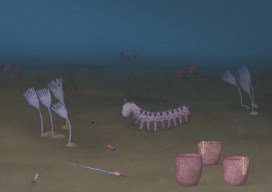
Once animated models of all 12 species were created we placed them in a realistic marine environment. Study of the rocks preserving the Chengjiang fossils suggests these animals lived in a relatively shallow, well-lit sea, perhaps 50 metres deep and characterised by a flat, muddy seafloor. A continuous shower of organic particles is thought to have filled the water column, as in modern oceans.
Reconstruction of the Cambrian seafloor with ‘marine snow’
Based on present-day marine ecosystems, we infer that the number of immobile suspension feeders would have been much greater than the number of predators. As a result, we included multiple individuals of the suspension feeders Cotyledion, Saetaspongiaand Xianguangia, which were tightly grouped together, but only a small number of the active predators Amplectobelua and Onychodictyon.
This scene is now populated with animals, including two predators: Amplectobelua (swimming) and Onychodictyon (centre)
The final step involved setting up a camera and user interface to allow visitors to discover the various animals in our interactive environment. For this we worked with creative digital consultancy Fish in a Bottle to identify eight locations, each focused on a different animal.
As the video above shows, users can navigate between locations by touching an icon on the screen, and when the Cambrian Diver sub arrives at a location information about the animal, its mode of life and its closest living relatives is presented on-screen. A physical joystick allows users a 360-degree rotation to look around the scene, and explore the ancient watery world.
This project was significantly bigger than the Out of the Deep work we had done previously with the Museum, mainly because of the complicated approval procedure needed for 20 individual 3D models. Along with three large illustrations, two animations and the interactive application this was a big workload! Fortunately, we managed to finish the whole project on time for the opening of the exhibition. – Martin Lisec
The latest display in our single-case Presenting… series takes a look at the famous Piltdown Man hoax, and Life Collections manager Mark Carnall tells us how the display came about…
Visiting researchers to the zoology collections at the Museum often give us an excuse to dig deeper into our own material, and one such recent enquiry led me into the intriguing story of the Piltdown Man hoax.
Professor Andrew Shortland from Cranfield University contacted us to enquire about the Piltdown Man material in our collections, as part of research for a book on hoaxes and forgeries in anthropology that he is writing with Professor Patrick Degryse of KU Leuven.
I knew we had some Piltdown material here thanks to this page written by Malgosia Nowak-Kemp, but I hadn’t had an excuse to investigate any further. The enquiry was also timely as we’d just transferred a collection of palaeoanthropology casts, models and reconstructions from our Earth collections to bring our human collections into one place. I knew from our move project team that there was some Piltdown material awaiting processing – perfect.
For those who don’t know the Piltdown Man story, a short history is in order. In the early 20th century, amateur fossil hunter Charles Dawson brought a collection of human remains excavated from gravel pits in Sussex to the attention of Arthur Smith Woodward, then Keeper of Geology at the British Museum (Natural History). Woodward and Dawson collected further material and presented the remains as those of Eoanthropus dawsoni (‘Dawson’s dawn man’), an important fossil human from Britain.
Group portrait of the Piltdown skull being examined. Back row (from left): F. O. Barlow, G. Elliot Smith, Charles Dawson, Arthur Smith Woodward. Front row: A. S. Underwood, Arthur Keith, W. P. Pycraft, and E. Ray Lankester. Charles Darwin looks on from a portrait on the wall. Image via Wikipedia.R.F. Damon-produced endocast and associated label recording the presentation of this specimen to the Museum by Arthur Smith Woodward
The discovery looked set to put Britain on the map when it came to evidence of human evolution, but suspicions were quickly raised about the authenticity of the material. Such was the skill of the forgery – meticulous breaking, abrading and staining of various archaeological and historic specimens – that it wasn’t until dating techniques, chemical analyses and some experimental palaeoanthropology in 1953 that the hoax was conclusively put to bed.
In turned out that the Piltdown ‘remains’ were a mix of medieval bone, an orangutan jaw, and chimpanzee teeth maltreated to look like an evolutionary intermediate between humans and other apes.
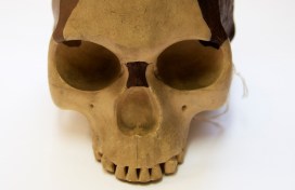
For 40 years or so the hoax refused to go away and numerous casts, models and reconstructions of Piltdown Man were made, sold, exchanged and gifted to museums and universities. These included casts of the original material as well as reconstructions of the skull and even reconstructions of the endocast – a cast of the inside of the skull.
The Museum has a selection of this material, but as Professor Shortland examined the collections, two specimens stood out.
The first is an R. F. Damon-produced endocast presented to the Museum by Arthur Smith Woodward himself. Smith Woodward was known as an expert on fossil fish but published widely on zoological topics. As a scientist of some repute there’s been long-standing speculation about his role in the hoax. Was he wholly duped by Dawson, or was he in on the hoax from the beginning? If it’s the former, then the presentation of this endocast shows Smith Woodward disseminating research he presumably took some pride in. If it’s the latter, perhaps it was a way of cementing the hoax as legitimate by spreading specimens far and wide.
Joseph Weiner’s experimental fake created by modifying an orangutan jaw, alongside a cast of the Piltdown jaw
The second significant specimen is a worked orangutan jaw produced by Joseph Weiner, one of the three authors who debunked the hoax in a 1953 Nature paper titled The Solution of The Piltdown Problem. Weiner modified the orangutan jaw to replicate the original hoax specimen. Thanks to Professor Shortland’s knowledge of the hoax, he sent through a copy of Weiner’s book on the Piltdown Man where this exact specimen is pictured.
The Piltdown Man hoax wasn’t the first and certainly won’t be the last hoax, fake or forgery in the history of science, but it remains one of the most well-known and stands as a warning of the dangers of hubris in the discovery and description of the natural world.
The Weiner jaw and Damon endocast will be on display alongside other Piltdown Man material in our Presenting… case from 9 January to 8 March 2020.
First Animals exhibition is on show until 24 February 2020
For our current exhibition, First Animals, we’ve taken this collaboration to a new level by commissioning original works from a total of 22 artists, all part of Oxford Printmakers Co-operative (OPC) – a group of over a hundred printmakers which has been running for more than 40 years.
First Animals looks at the very earliest evidence of life on Earth, dating back half a billion years. Some of the fossils on display are shallow impressions in the rock – the only direct evidence we have that life existed at that time.
Amplectobelua symbrachiata – one of the incredible Cambrian fossils from the Chengjiang site in China
To kick-start the project we ran a series of workshops for OPC artists to meet the Museum researchers working on the exhibition, and to see the fossils first hand. There were also opportunities to draw directly from these unique fossils, many of which have never been displayed in the UK before.
Discussions between researchers and artists revealed fascinating similarities between these ancient fossils and the process of printmaking. Sally Levell, of Oxford Printmakers Co-operative, explains:
I was completely fascinated by the fossil collection in the Museum, especially the fine specimens from Chengjiang and Newfoundland. They are preserved as mere impressions in the rock, so they are, in essence, nature’s prints.
Each printmaker partnered with a researcher who could answer questions, provide extra info and help the artist decide which specimen or subject to depict in their final print. It’s clear from talking to the printmakers that this direct contact with the experts was invaluable and made the work really meaningful.
Xianguangia by Charlie Davies
We couldn’t have worked without the patient explanations and “show and tell” sessions with the three main researchers – Dr Jack Matthews, Dr Imran Rahman and Dr Duncan Murdock. They were just excellent and their dedication to their work was an inspiration to all of us printmakers.
Sally Levell
Over a period of around seven months, ideas blossomed and printing presses were put into action, with the printmakers exploring the forms, textures and evolution of the fascinating first animals. The final result is First Impressions, an enticing art trail of twenty-five prints dotted around the Museum, both within the First Animals exhibition gallery and nestled within the permanent displays.
Ottoia by Jackie Conway
Such a large group of artists brings a huge variety of techniques and styles, all under the umbrella of printmaking; from a bright, bold screen print in the style of Andy Warhol, to a delicate collagraph created from decayed cabbage leaves! To take part in the art trail yourself, simply grab a trail map when you’re next in the Museum.
Workshop printers inking up their plates
But our foray into fossils and printmaking didn’t stop there. OPC member Rahima Kenner ran a one-day workshop at the Museum where participants made their own intaglio prints inspired by the First Animals fossils. The group of eight people featured artists and scientists alike, all keen to capture the unique fossils through print techniques.
Designs were scratched onto acrylic plates and inked up, before a professional printing press created striking pieces to take home. Participants also explored techniques such as Chine-Collé, the addition of small pieces of paper to create texture and colour underneath the print.
It was a delight to be able to share with the group our enthusiasm for these discoveries in the medium of making the drypoint prints and to share their enjoyment of learning and using the new techniques. Some lovely work was produced in a single day.
Rahima Kenner
A plate about to go into the pressA finished print, using intaglio and chine-colle
The First Impressions project has been transformative for the Museum team and for the Oxford Printmakers Co-operative. Catriona Brodribb describes its impact on the printmakers :
It’s been a great opportunity to challenge one’s own artistic boundaries in terms of stretching the imagination, and for our members to throw themselves into something new, and enjoy responding to such ancient material in a contemporary way.
The First Animals and First Impressions exhibitions are open until 24 February 2020 and are free to visit.
Over the past few months our researchers have been working with University of Plymouth illustration student Abigail Harris, who has delved into the weird and wonderful world of some of the earliest animals. Here, Abigail tells us about the process that led to the creation of her Cambrian artwork, inspired by our First Animals exhibition.
I first visited the Museum in April this year when I was given the opportunity to collaborate with scientists as part of a module in my BA in at the University of Plymouth. Things kicked off with a short talk about the Ediacaran and Cambrian geological periods, when Earth’s first animal life started to appear.
I quickly narrowed my interest down to fossils from the Cambrian period which are more complex life forms, more similar to life today. A collection of small fossils from the Chengjiang fossil site in Yunnan province, China was the inspiration for some initial observational drawings.
A sketchbook page showing initial sketches and observations of OnychodictyonFinal illustration of Cotyledion
After returning to Plymouth University, I began to develop these initial sketches and observations, continuing to research the Chengjiang material and learning more about the characteristics of some of the creatures preserved as fossils.
I wanted to create an under-the-sea ecology reconstruction showing a diversity of life forms, focusing on Onychodictyon, Cotyledion, Cricocosmia, Luolishania, and Paradiagoniella.
A five-step process was used for each reconstruction. Initially, I would sketch the fossil as I saw it, then I would research the characteristics and features of that animal, making a list of things to include in my drawing. A second drawing would then include all of these characteristics, not just what was initially visible in the fossil.
These rough sketches were then sent to the scientists for feedback, helping me to redraw and paint the illustrations with watercolour, before scanning and digitally editing each painting. Lastly, I created a background and added my illustrations.
Initial under under the sea ecology reconstruction.
Although the reconstructions were not completely finished by the time of my project deadline, I returned to the Museum in July and was given a tour of the First Animals exhibition by Deputy Head of Research Imran Rahman, as well as the opportunity to discuss how to improve my artworks for accuracy.
Another round of sketching and painting led to the final piece, shown at the start of this article, complete with an added digital background of the seafloor, and darkened to reflect the murky world of a Cambrian ocean, 50 metres below the surface.
The latest exhibition in our Contemporary Science and Society series, First Animals, tells the tale of Earth’s mysterious early animals, which evolved in the sea over half a billion years ago. Here, Dr Imran Rahman, Deputy Head of Research at the Museum, introduces some of the fossils that form a key part of this story.
From sponges to sea slugs and hagfish to humans, all animals alive today trace their roots back to a common ancestor that lived in the ocean more than 600 million years ago. We have no direct evidence of this first animal, but the fossil record reveals some of its earliest descendants. Our First Animals exhibition explores the evidence for Earth’s earliest animal life, attempting to answer the ‘what’, ‘when’, ‘how’ and ‘why’ of the origin of animals.
Yunnanozoon lividum from the Chengjiang fossil site had a long body with several filament-covered arches at the front and a fin-shaped structure towards the back. It cannot be confidently assigned to any known animal group.
First Animals features the oldest animals yet recovered from the fossil record, including specimens from 571-million-year-old rocks in Newfoundland, Canada. These represent the remains of originally entirely soft-bodied organisms, which have proven difficult to classify because they look so different to living species. However, new research on their anatomy and how they grew, including work by Museum researcher Dr Frankie Dunn, suggests they were early animals.
Charnia masoni consisted of alternating branches arranged along a frond. It is thought to be one of the oldest animal fossils yet found.
Microscopic fossils record the first animal skeletons, which first appeared about 550 million years ago. These include the remains of complete animals, as well as fragments such as spines and scales. Work by Museum researcher Dr Duncan Murdock using a particle accelerator to generate X-ray images of these tiny fossils has allowed us to reconstruct how the skeletons changed as they grew. This helps to establish the modern groups to which these ancient animals belonged, and unravels the mystery of why animals evolved hard skeletons when they did.
Virtual cross-sections through small shelly fossils created using X-ray imaging.
The most complete evidence for the early evolution of animals comes from sites of exceptionally-preserved fossils, or Lagerstätten, which retain impressions of soft tissue as well as hard parts, and include rare soft-bodied animals like worms and jellyfish.
First Animals brings together extraordinary specimens from three key fossil sites: Sirius Passet in northern Greenland (518 million years old), Chengjiang in Yunnan province, China (518 million years old) and Burgess Shale in British Columbia, Canada (508 million years old). This includes 55 unique fossils loaned by Yunnan University in China, as well as specimens from the University of Bristol and the Royal Ontario Museum.
The mollusc Halkieria evangelista from the Sirius Passet fossil site had a long body covered in hundreds of overlapping hard plates, with a large shell plate at either end.The arthropod Haikoucaris ercaiensis from the Chengjiang fossil site had a semicircular head shield with a pair of large grasping appendages, a segmented body and a short tail.The worm Ottoia prolifica from the Burgess Shale fossil site had a spiny proboscis and a long trunk that was divided into a series of fine rings.
These exceptionally-preserved fossils reveal the evolutionary diversification of life during the so-called ‘Cambrian explosion’. Through careful study of the fossils, scientists have begun to reconstruct the very first animal ecosystems, which are brought to life in the exhibition through a series of stunning digital reconstructions and the Cambrian Diver interactive installation. This allows visitors to explore a 360-degree oceanic environment in a virtual submersible craft, coming face-to-face with some of the first animals on Earth!
Digital reconstruction of the sea floor 518 million years ago, based on specimens from the Chengjiang fossil site, Yunnan province, China.
Video by Mighty Fossils.