Lynn Margulis and the origins of multicellular life

To mark International Women’s Day Professor Judith Armitage, lead scientist on the Bacterial World exhibition, reflects on the ground-breaking – and controversial – work of evolutionary biologist Lynn Margulis

Iconoclastic, vivacious, intuitive, gregarious, insatiably and omnivorously curious, partisan, bighearted, fiercely protective of friends and family, mischievous, and a passionate advocate of the small and overlooked.

Lynn Margulis at the III Congress about Scientific Vulgarization in La Coruña, Spain, on November 9, 2005. Image: Jpedreira, CC BY-SA 2.5

These are all words used to describe evolutionary biologist and public intellectual Lynn Margulis. Intellectually precocious, Margulis got her first degree from the University of Chicago aged 19, but it was her exposure to an idea about the evolution of a certain type of cell that ignited a lifelong focus of her work.

This idea claimed that eukaryotic cells – cells with a nucleus, found in all plants and animals, but not bacteria – were first formed billions of years ago when one single-celled organism – a prokaryote – engulfed another to create a new type of cell. This theory, known as endosymbiosis, was laid down in a paper by Margulis in 1967. It brought her into conflict with others, including the so-called neo-Darwinists who believed in slow step-wise evolution driven by competition between organisms, not cooperation.

So what happened in the earliest evolution of these crucial cells? Initially, one bacterium ate a different, oxygen-using bacterium but didn’t digest it. Over time the two became interdependent and the bacterium took over almost all of the energy-generating processes of the host cell, becoming what we now call a mitochondrion. This allowed the cell to evolve into bigger cells and eventually form communities and develop into multicellular organisms.

Animal cells evolved when one single cell, possibly an archaeon, engulfed an aerobic bacterium – one that used oxygen to release energy. The bacterium evolved into the mitochondrion, the powerhouse of the cells of humans and other animals. A similar process created the chloroplasts found in plant cells.

These early mitochondria-containing organisms continued to eat other bacteria, and on more than one occasion they ate a photosynthesising cyanobacterium which evolved into a chloroplast, a structure now found inside plant cells.

The revolution in DNA sequencing that started in the 1970s, and continues today, eventually vindicated Margulis’ position on this ancient sequence of events. It revealed that chloroplasts and mitochondria both contain DNA with the same ancestry as cyanobacteria and proteobacteria respectively. In other words, both chloroplasts and mitochondria have evolved from ancient bacteria.

Margulis’ enthusiastic support for these ideas led her to think about the role of biology in the geology of Earth and some of its major changes, in particular the oxygenation of the atmosphere by cyanobacteria around 2.5 billion years ago. Mitochondria use oxygen, and so must have evolved from bacterial ancestors that arose after the cyanobacteria started to produce oxygen through photosynthesis.

Margulis met Gaia theorist James Lovelock soon after her seminal publication on endosymbiosis. At the time, Lovelock was looking at the composition of the atmosphere and factors causing change, including oxygen levels. He was starting to think of the Earth as a system – Gaia as it became known – where the planetary environment is regulated and kept stable by biological activity.

This meeting brought together two scientific outliers. Together they produced highly controversial articles on the “atmosphere as a biological contrivance”. Lovelock believed in concentrating on examining the systems as they are now, while Margulis brought deep time and evolutionary depth into the picture.

Margulis’ ideas were not always right, and she was enormously controversial in her time, but she made people think again. And in doing so she moved our understanding of things as apparently academically distant as the evolution of tiny cells billions of years ago to the stability of Earth’s environment today.

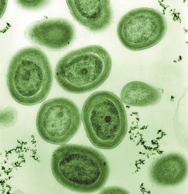
Top image: Euglena, a single cell eukaryotic. By Deuterostome [CC BY-SA 3.0]

Bacteria that changed the world: Prochlorococcus

In our Bacterial World exhibition we offer a selection of ten bacteria that have changed the world, some in profound ways. In this series of short fact-file posts we present one of the ten each week. This week’s bacteria are…

Prochlorococcus
– the Oxygen-Makers

Where they live
Prochlorococcus bacteria grow anywhere damp, in salt water or fresh. They are similar to the blue cyanobacteria which thrived in the far-distant past on Earth.

Why they are important
2.3-2.4 billion years ago, cyanobacteria in the oceans began producing oxygen for the first time, changing the Earth’s environment completely.

How they are named
The Greek word for blue is cyan, giving the blue cyanobacteria their name. Until recently, they were known as blue-green algae, but cyanobacteria are actually an earlier and simpler form of life than algae.

How they work
Like all cyanobacteria, Prochlorococcus bacteria harvest energy from the Sun, absorb carbon dioxide and give out oxygen – the process called photosynthesis.

Top image: Transmission Electron Micrograph (TEM) image of Prochlorococcus coloured green
Copyright: Luke Thompson, Chisholm Lab; Nikki Watson, Whitehead (MIT), 2007

Bacterial Girl


We couldn’t resist. The moment we came up with the title of our special exhibition, Bacterial World, we were all humming Madonna’s 1985 hit. So here it is – a bacteria-themed version of Material Girl – written, performed and illustrated by the talented Museum team.

In place of “cold hard cash”, you’ll learn that bacteria were involved in the creation of life on Earth, and you’ll find DNA exchange and photosynthesis in place of kisses and hugs. Have a listen… and try to stop yourself dancing.

Of course, you’ll need the full lyrics to sing it in your bedroom with a hairbrush:

BACTERIAL GIRL

Some bugs make you feel unwell
And we’ve all heard of them
But look inside and you will find
That bacteria are your friend

They’ve been around since way back when
In the ocean life began
But nowadays they’re everywhere
So I think you’ll understand, that we are…

Living in a bacterial world
And I am a bacterial girl
You know that we are
Living in a bacterial world
And I am a bacterial girl

They spent some time in the sun
Began to photosynthesise
Put oxygen in the air we breathe
I’m telling you no lies

Now E. coli’s got a real bad rep
For causing people pain
But what you got to realise
Is it’s only one bad strain

’cause we are
Living in a bacterial world
And I am a bacterial girl
You know that we are
Living in a bacterial world
And I am a bacterial girl

Now some bugs love to snuggle up to
Exchange their DNA
Other cells are armed with spears
That wipe enemies away

Resistance to our medicines
You could call it evolution
But microbes might just hold the key
To a medical solution, ’cause we are

Living in a bacterial world
And I am a bacterial girl
You know that we are
Living in a bacterial world
And I am a bacterial girl

You know that we are

Living in a bacterial world
And I am a bacterial girl!

Bacterial World is open until 28 May 2019.

Credits for this little bit of brilliance go to:
Vocals, violin: Laura Ashby
Words, banjo, guitar, recording: Scott Billings
Illustrations: Chris Jarvis

Presenting… Christmas Island

By Eileen Westwig, Collections Manager in the Museum’s Life Collections.

About 320 km south of Java in the Indian Ocean lies Christmas Island. Although discovered and named on Christmas Day in 1643, the island remained unexplored until its first settlement in 1888, a development which had dire consequences for some of its native species.

Christmas Island is home to a variety of endemic animals such as rats, land crabs, butterflies and many birds. The accumulation of bird droppings over thousands of years made the island rich in phosphate, and the commercial potential of these deposits brought many expeditions to the island. With the ships’ cargo came black rats.

Two species of endemic rats, Maclear’s Rat (Rattus macleari) and the Bulldog Rat (Rattus nativitatis) went extinct within 20 years of settlement, despite having been previously very numerous on the island.

One of the skins of Maclear’s Rat (Rattus macleari) collected by H.E. Durham, and now held in the Life Collections of the Oxford University Museum of Natural History.

Maclear’s Rat, seen at the top of the page in an illustration from an 1887 publication, was described as chestnut brown above, with a partly white, long tail. It was once the most numerous mammal on the island ‘occurring in swarms’. The Bulldog Rat had a much shorter tail and a layer of subcutaneous fat up to 2 centimetres thick, the function of which is unknown to this day.

The likely cause of their extinction was the introduction of diseases by the ship rats, to which the Christmas Island rodents had no immunity. The disappearance of the native rats also had a knock-on effect: the parasitic Christmas Island Flea (Xenopsylla nesiotes) depended on the rats as hosts, and so the fleas became extinct with the rats’ demise.

In 1901 Dr. Herbert E. Durham, a British parasitologist investigating the origins of beriberi disease, led an expedition to Christmas Island. During his visit he collected several specimens of Maclear’s Rat, but was unable to find any Bulldog Rats, despite a lengthy search and the offer of a reward. Two of the nine Maclear’s Rats Durham obtained showed abundant parasites, trypanosomes, in their blood.

Christmas Island possesses quite a number of peculiar species in its fauna, and it is regrettable that observations were not made before animals had been imported to this isolated station, as well as that my own notes are so incomplete.

Dr. Herbert E. Durham

Durham also found blood parasites in the native fruit bats (Pteropus melanotus) but noted that these were unlikely to have been introduced, instead were “an old standing native occurrence.” These bats still inhabit various islands in the Indian Ocean, including Christmas Island, where they are critically endangered.

Original letter by H.E. Durham offering his Christmas Island specimens to the Museum in 1938.

The Museum holds a range of material from Christmas Island, including six skins and three skulls of Rattus macleari, which were collected by H. E. Durham in 1901-02, and donated in 1938.

Visit the Museum’s Presenting… case between now and 6 March to see Christmas Island specimens from the collections.

Precision antibiotics – the future treatment of infections?

by Hannah Behrens

In our Bacterial World Science Short event series, researchers present their latest findings related to themes in the exhibition. At a recent Science Short, Hannah Behrens, a University of Oxford PhD student, explained how bacteria become resistant to antibiotics and how the species-specific antibiotics she studies might reduce the worrying rise in antimicrobial resistance.

Bacteria that are resistant to antibiotics present a huge problem. I work on developing new antibiotics that will slow the development of bacterial resistance.

But let’s not get ahead of ourselves. Your body is full of bacteria. In fact, there are more bacteria than human cells in your body. Most of these bacteria are good for you; they help you digest food and protect you from diseases.

But once in a while a harmful bacterium causes an infection. This could be a lung, wound, or bladder infection, or something with a fancy name like, Black Death, tuberculosis, leprosy, syphilis or chlamydia. The doctor will then prescribe you antibiotics to kill the offending bacteria.

Hannah Behrens delivers her Science Short talk at the Museum

The development of antibiotics in the 20th century was a major breakthrough. For the first time bacterial infections could be effectively and rapidly treated. Since 1942, when antibiotics first became available, we have discovered many new antibiotics which have saved millions of lives.

However, in the last 30 years we have not managed to develop any new antibiotics. During the same time, many bacteria have adapted to become resistant to the antibiotics we do have. In 2017, a woman in the US died because she had an infection with bacteria that were resistant to all available antibiotics. It is estimated that already 700,000 people in Europe alone die because of resistant bacteria per year. What is happening?

Bacteria are forming a lawn on this plate (light areas); where an antibiotic has been spotted on the bacteria they die and leave the surface blank (dark areas).

Every time we treat bacteria with antibiotics, most die, yet a few resistant bacterial cells survive. Like Rudolph the red nosed reindeer, the resistant bacteria are usually at a disadvantage until a special situation arises (a foggy night for Rudolph; treatment with antibiotics for resistant bacteria).

Under usual circumstances, producing a resistance mechanism is a disadvantage: it wastes energy and slows down growth, so very few bacteria are resistant. Only when all the non-resistant bacteria are killed by antibiotics do the resistant ones thrive. They have no more competition, and have all the resources, such as food and space, to themselves.

The more we use antibiotics, the more resistant bacteria we get. It is essential not to use antibiotics carelessly.

More antibiotics are used in animal farming than on humans. If we eat less meat, and so reduce the farming of livestock for food, we may reduce the growth of resistance bacteria. Another approach is to only take antibiotics when the doctor prescribes them. Antibiotics do not help against viral infections like colds. In many low and middle income countries, antibiotics are available in supermarkets and it is no coincidence that these countries have higher levels of resistant bacteria.

The precision antibiotics research group in the Department of Biochemistry at the University of Oxford

Apart from avoiding the unnecessary use of antibiotics, scientists – including me – are trying to develop better therapies against bacteria. I study precision antibiotics: drugs that specifically kill one species of bacteria. The advantage of this is that all good bacteria remain unharmed and only the disease-causing species is targeted. This also means that only resistant bacteria from this one species get an advantage to thrive.

I am interested in species-specific antibiotics against Pseudomonas aeruginosa. This bacterial species causes lung and wound infections and, according to the World Health Organization, is one of the three bacteria for which we most urgently need new antibiotics. Colleagues of mine tested different precision antibiotics against Pseudomonas and found one that is better than the others, called Pyocin S5.

Hannah’s painting of how researchers think pyocin antibiotics kill bacteria. The pink bacterium produces pyocins (pink balls), which enter the susceptible blue bacteria through pores (blue). The blue bacteria mistake the antibiotic for a nutrient and open the pore to let it in. Once inside the bacterium it forms a pore in the inner membrane which causes leakage of the cell contents and kills the cell.

I am now investigating how stable this antibiotic is, how it recognises this specific species of bacteria and how it enters the bacterial cells. This knowledge is important to decide on how to store, transport and administer the drug. I also hope that understanding why Pyocin S5 is more effective than the other antibiotics will allow us to design more effective, targeted antibiotics in the future.

My hope is that one day we will treat all bacterial infections with precision antibiotics and that antibiotic resistance will become a problem of the past.

Bacteria: captured and cultured

For our new exhibition, Bacterial World, we embarked an exciting science/art experiment to make visible the colonies of bacteria present on a wide range of our everyday items and belongings. Once cultured and photographed, eight of these colonies were captured by artist Elin Thomas as a set of crochet artworks that are on display in the exhibition. Our exhibitions officer Kelly Richards tells us more…

For every human cell in your body, a bacterial cell is also present. These bacteria are part of our microbiome, a vast array of microorganisms that use our body as a home and our food as a source of nutrients. In return, the bacteria help us to digest food, maintain our immune systems and keep dangerous bacteria at bay. In fact without these bacteria we would be very sick indeed.

It’s hard to see our microbiome because individual bacteria can easily be as small as 0.2 microns; you could fit over a thousand of these smallest bacteria on one side of a red blood cell. But if we can select and artificially grow the bacteria, their colonies become living, breathing cities visible to the naked eye.

Click the images above to find out more about culturing bacterial colonies

Colonies, both natural and artificial, can contain billions of bacteria as well as the materials that they secrete such as slime, which helps them to move across surfaces, and antibiotics, which kill off other bacterial colonies that could compete for food and space. In their attempts to dominate the space and food available, as well as get enough oxygen to live, colonies can create beautiful, complex structures.

We had a go at visualising the bacteria that live invisibly alongside us by asking visitors to take part in a simple experiment. With the help of microbiologist Rachael Wilkinson, we took items such as coins, keys and jewellery and touched them lightly against agar plates – dishes containing a nutrient-rich jelly that aids bacterial growth. The agar plates were then given to Nicole Stoesser, a clinical microbiologist at the John Radcliffe Hospital, who grew them in the safe environment of the laboratory.

Many different types of colonies grew from the objects we printed. In the collage above, eight of these colonies have been represented as crocheted Petri dishes by artist Elin Thomas. These artworks are on display in the Bacterial World exhibition until 28 May 2019.

In the gallery below is a photograph of every participant’s plate, whether anything grew in it or not. Click on an image to see a larger version. If you took part in the experiment you will be able to identify your own plate from its number.

The results go to show that we really are living in a bacterial world!