A tale of two seahorses

Real or fake? Do replicas have a value of their own? Elaine Charwat is exploring this in her PhD, using the Museum’s large collection of natural history models and casts to research their role in science. Here she tells the story of the fascinating fish that caught her imagination…

By Elaine Charwat

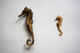
It all started with a seahorse. Last year, I walked into a little seaside shop, and I spotted a seahorse. I instantly flipped back to the happy day I bought my first dried seahorse as a child, the beginning of a life-long passion for the natural world. The man behind the counter smiled: “It’s a fake.” Really? “3D printed.” It looked absolutely perfect. Tracing its lines with my fingers, I said, “It’s a model”.

Ever since I became interested in models and replications, I have encountered this perception of them as “fakes”. Quite recently, I heard the curator of a natural history museum call the cast of a dinosaur skeleton a “fake”. Models in natural history – and in this I include casts and reproductions – are what the Germans call “Wissensdinge”, objects that contain, distribute and generate knowledge. In this aspect, the real specimen and the model meet. Models are made from a vast array of materials with often astonishing skill and technologies. They represent what we know about a particular organism at a certain point in time. They have a history, a context.

Long live the replica! Most of our most beloved dinosaur skeletons in museums are partly or fully casts of bones, like Stan’s here at the Museum. Almost complete skeletons like Stan’s are extremely rare, and casts allow us to share and preserve them. Accompanying models give the bones “flesh and blood” – and provide a snapshot of what was known about the dinosaur when the model was made.

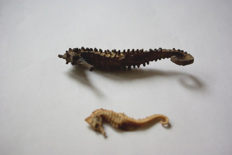
But they are also ambassadors, and this is something I realised when I held the “fake” 3D-printed seahorse in my hand. While it becomes ethically problematic to buy specimens of organisms like seahorses, something of it is captured, and communicated, in a reproduction. I can still trace its exoskeleton, and marvel at its strange symmetry. This symmetry, incidentally, is being analysed for its potential in robotics. Seahorses have unusual tails – instead of the cylindrical trail structure found in most animals, theirs have a square cross-sectional architecture, resulting in a unique combination of toughness and flexibility. In fact, when studying the unique abilities of the seahorse’s tail, researchers have actually used 3D-printed specimens.

Seahorse from the Museum’s collection. Even in Victorian times, long before 3D printers, there seems to have been a desire to emphasise that souvenir seahorses were “natural” – i.e. not man-made. Was it because seahorses are easily preserved and so attractive when dead and dried?

The Oxford University Museum of Natural History has a largely unexplored wealth of models and casts. Many of them date to the second half of the 19th Century, the heyday of their production. Made from glass, wax, metal, wood, plaster, papier-mâché or, indeed, actual bone and feathers, they were modelled, cast, sculpted, glued, painted and mounted to enhance and preserve our understanding and appreciation of nature. But they also tell of scientific discoveries and controversies, research and teaching, rivalries and collaboration, politics and society, ideas and identities.

Small IMGP5899
Spot the replica – both the specimen and the 3D printed seahorse are “Wissensdinge”, they have a context and provide valuable information.

I will trace these complex relationships in a collaborative and interdisciplinary PhD project called “Nature of Replication”. This is funded by the AHRC and jointly supervised by the Institute of Archaeology, University College London, and the Oxford University Museum of Natural History.

The 3D-printed seahorse now lives alongside my real seahorse. So I like to think of my project as a journey that started with one seahorse, and continues with another.

Understanding beeswax

By Tuuli Kasso, PhD in Science Fellow at the Natural History Museum of Denmark, University of Copenhagen. Tuuli is a visiting researcher, who has used the Museum’s collection to help her understanding of beeswax. 

When working on the dissertation for my MSc in Archaeological Science last year, I explored the medieval craftsmanship of sealing wax. I was interested in the way the medieval wax seals had flaked, as the beeswax dried out. Drawing on my previous education in conservation techniques, I began a close investigation of the prestigious material, beeswax.

Medieval craftsmen used a range of dangerous materials to make sealing wax. The red pigment cinnabar, a mercury (II) sulphide, and red lead, are now known to be extremely poisonous.

Although some of the ingredients of sealing wax are very hazardous, there is nothing dangerous in beeswax… except the bees! Produced by honey bees, Apis mellifera, honey and beeswax were important commodities in the Middle Ages. Beekeeping was a skilful profession, housing colonies in woven hives, known as skeps. Colonies were carefully selected to overwinter for the next season.

Manuscript illuminations provide detailed information on the types and construction of beehives in the Middle Ages.England, 13th century. British Library Royal 12 C XIX f. 45.

Beeswax was also important in the Middle Ages for lighting, and beeswax candles were preferred for their pleasant smell. After the Protestant Reformation in the 16th and 17th centuries, the religious use of candles decreased, so demand for beeswax declined.

Even today, the Catholic and Orthodox Churches still require the candles they use to contain a proportion of beeswax.

On my quest to understand the degradation of beeswax in sealing wax and write my disseration, I was very lucky to use some samples from the entomological collections from the Oxford University Museum of Natural History. After some early mornings spent amongst the Westwood collection, I found the perfect specimens of natural honeycombs, from the 19th century. The old hand-written labels were also a lovely encounter when exploring the historical collections.

I compared the samples to modern beeswax and medieval seal samples, and learned that the degradation of beeswax is caused by multiple factors, triggered also by storage conditions. The composition of beeswax is very complex, and there are differences caused by the age of the bee in addition to geographical provenance.

A selection of bee specimens from the Museum’s collection.

The recent catastrophic decline of bee populations has drawn focus to save the bees, and in my PhD research (University of Copenhagen and University of Cambridge) I will explore the recovery of ancient DNA and proteins of bees from beeswax, to cast light on the health of bee populations over time.

Bursting into life

By Ricardo Pérez-de la Fuente, Museum Research Fellow

One of the earliest and toughest trials that all organisms face is birth. In egg-laying animals, the egg shell that has protected the embryo during its early development ultimately becomes a hard barrier between the animal and its life out in the world. The bursting of the egg is literally a threshold moment, and there are many ways to crack an egg…

Some animals break the egg membranes using dissolving chemicals; others physically, mechanically tear their way through the shells. Among the latter, a great diversity of animals use specialised devices called egg bursters. These vary greatly among the many arthropods and vertebrates that use them, but perhaps the most famous example is the ‘egg tooth’ that is present on the beak of newborn chicks.

Four complete Tragychrysa ovoruptora newborns preserved together with egg shell remains and one egg burster. Modified from the open access Palaeontology paper.

My colleagues and I have found an exceptional fossil in 130 million-year-old Lebanese amber. Inside, trapped together are newborn larvae from Green Lacewings, the split egg shells from where they hatched, and the minute egg bursters that the hatchlings used to crack the egg. This is a first: no definitive evidence of these specialised egg-bursting structures had been reported from the fossil record of any egg-laying animals, until now.

The finding has been recently published as open access in the journal Palaeontology. Because multiple newborns were ensnared and entombed in the resin simultaneously, the fossil larvae have been described as the new species Tragichrysa ovoruptora, meaning ‘tragic green lacewing’ and ‘egg breaking’. A sad event, indeed, taking place in an ordinary day 130 million years ago in the Cretaceous forests of Lebanon, yet a happy circumstance now that we can take a privileged glimpse into the adaptations and behaviours of these fascinating tiny creatures.

The hatchlings from modern Green Lacewings open a slit on the egg with a ‘mask’ bearing a saw-like blade. Once used, this ‘mask’ is shed together with the embryonic cuticle and is left attached to the empty egg shell.

With the help of Amoret Spooner, Collections Manager at the Museum, egg clutches from modern green lacewings were found in the Museum collections. These eggs happened to have the intact egg bursters still attached to them, and proved to be crucial to understand that we had the same structures preserved in the amber together with the newborn larvae.

Two Tragychrysa ovoruptora newborns preserved together with egg shell remains and two visible egg bursters (right inset). Modified from the open access Palaeontology paper.

Green Lacewing larvae are small predators that often carry debris as camouflage, using their sickle-shaped jaws to pierce and suck the fluids of their prey. Whereas the larvae trapped in amber differ significantly from modern-day relatives, in that they possess long tubes instead of clubs or bumps for holding debris, the studied egg shells and egg bursters are remarkably similar to those of today’s green lacewings.

The larvae were almost certainly trapped by resin while clutching the eggs from which they had freshly emerged. Such behaviour is common among modern relatives while their body hardens and their predatory jaws become functional. Indeed, the two mouthparts forming the jaws are not assembled in most of the fossil larvae, which indicates, together with the large relative size of the head and legs, that they were recently born.

Detail of a head with the jaws still dislodged, indicating that the larva was recently hatched when it was ensnared by amber and the jaws had not yet had time to fully assemble.

It may seem reasonable to assume that traits controlling a life event as decisive as hatching would have remained largely unchanged during evolution. In fact, we see in very closely related insect groups different means of hatching that can entail the loss of the egg bursters. So the persistence of a hatching mechanism in a given animal lineage through deep time can’t be determined without direct proof from the fossil record.

Reconstruction of two Tragichrysa ovoruptora newborns clutching the eggs from where they recently hatched, moments before they were trapped by resin. Larvae colour and egg stalks are conjectural. Extracted from the open access Palaeontology paper.

This new discovery shows that the mechanism green lacewings use to crack the egg was already established 130 million years ago. Overall, it represents the first direct evidence of how insects hatched in deep time, egg-bursting their way through into life.

*

The hatching mechanism of 130-million-year-old insects: an association of neonates, egg shells and egg bursters in Lebanese amber by Ricardo Pérez-de la Fuente, Michael S. Engel, Dany Azar and Enrique Peñalver is published as open access in Palaeontology this month.

Imagining lost worlds

Earlier this year University of Plymouth illustration student Rachel Simpson teamed up with our research fellow Jack Matthews to ‘bring the oldest multi-cellular organisms back to life’. Rachel tells us about the process of working with some of the most ancient fossil material and reveals the results of her illustrations and modelling.

Illustration by Rachel Simpson, created in collaboration with the Museum

In August 2018 I was lucky enough to travel to Newfoundland, Canada with Dr Jack Matthews to learn about and illustrate some of the extraordinary fossils found there. A highlight of the trip was going down onto the fossil surface – known as the MUN surface – to look at examples of organisms such as Beothukis, Charnia and Primocandelabrum, all of which date from the Ediacaran period, over 550 million years ago.

The MUN surface is the location of the fossils that I had worked on for my university project. I had spent the previous months sketching, drawing and bringing these organisms back to life from silicon casts, so it was amazing to be able to see the real specimens in situ and to sketch from the fossil surface.

Sketching directly from the fossils also provided a new challenge as I was unable to control factors such as the lighting, which is crucial to seeing the fossils clearly. Nonetheless, I learnt a lot about drawing on location.

Sketching at the fossil surface

While visiting Port Union I was able to use some of the old printing presses held by the Sir William F. Coaker Heritage Foundation to create work inspired by the fossils I had seen in the surrounding area. I love using printmaking in my own illustrative practice so it was a great experience to get to use these old presses (image at top of article).

We also had the chance to give a radio interview and talk to the Port Union community about the work that Jack and I had done, showing how science and art can work together.

On my last day in Port Union I was invited by a local potter to make some ceramic representations of the fossils I had been drawing there. I created models of Fractofusus and Aspidella, and discovered that re-imagining something in three dimensions is a very different process to recreating it as a drawing.

Rachel created ceramic representations of some of the Ediacaran organisms

For the final three days of the trip we relocated from Port Union to Trepassey to visit the Mistaken Point UNESCO World Heritage Site. Here, I saw the highly preserved Fractofusus specimens and made some more sketches. Using a small hand lens I was able to draw all the details that are invisible to the naked eye.

Using a hand lens allowed Rachel to pick out details in the Fractofusus fossil

Drawing on location in Canada provided a better idea of the organisms in relation to other surrounding organisms, something that is more obscure when working from museum specimens. This definitely informed my practice and meant that artwork created after the trip was more representative of the science.

When I returned to England, I created some new prints inspired by my time in Newfoundland, the fossils that I saw, and the printing process I was able to use in Port Union.

A set of prints made by Rachel based on her work in Newfoundland

Precision antibiotics – the future treatment of infections?

by Hannah Behrens

In our Bacterial World Science Short event series, researchers present their latest findings related to themes in the exhibition. At a recent Science Short, Hannah Behrens, a University of Oxford PhD student, explained how bacteria become resistant to antibiotics and how the species-specific antibiotics she studies might reduce the worrying rise in antimicrobial resistance.

Bacteria that are resistant to antibiotics present a huge problem. I work on developing new antibiotics that will slow the development of bacterial resistance.

But let’s not get ahead of ourselves. Your body is full of bacteria. In fact, there are more bacteria than human cells in your body. Most of these bacteria are good for you; they help you digest food and protect you from diseases.

But once in a while a harmful bacterium causes an infection. This could be a lung, wound, or bladder infection, or something with a fancy name like, Black Death, tuberculosis, leprosy, syphilis or chlamydia. The doctor will then prescribe you antibiotics to kill the offending bacteria.

Hannah Behrens delivers her Science Short talk at the Museum

The development of antibiotics in the 20th century was a major breakthrough. For the first time bacterial infections could be effectively and rapidly treated. Since 1942, when antibiotics first became available, we have discovered many new antibiotics which have saved millions of lives.

However, in the last 30 years we have not managed to develop any new antibiotics. During the same time, many bacteria have adapted to become resistant to the antibiotics we do have. In 2017, a woman in the US died because she had an infection with bacteria that were resistant to all available antibiotics. It is estimated that already 700,000 people in Europe alone die because of resistant bacteria per year. What is happening?

Bacteria are forming a lawn on this plate (light areas); where an antibiotic has been spotted on the bacteria they die and leave the surface blank (dark areas).

Every time we treat bacteria with antibiotics, most die, yet a few resistant bacterial cells survive. Like Rudolph the red nosed reindeer, the resistant bacteria are usually at a disadvantage until a special situation arises (a foggy night for Rudolph; treatment with antibiotics for resistant bacteria).

Under usual circumstances, producing a resistance mechanism is a disadvantage: it wastes energy and slows down growth, so very few bacteria are resistant. Only when all the non-resistant bacteria are killed by antibiotics do the resistant ones thrive. They have no more competition, and have all the resources, such as food and space, to themselves.

The more we use antibiotics, the more resistant bacteria we get. It is essential not to use antibiotics carelessly.

More antibiotics are used in animal farming than on humans. If we eat less meat, and so reduce the farming of livestock for food, we may reduce the growth of resistance bacteria. Another approach is to only take antibiotics when the doctor prescribes them. Antibiotics do not help against viral infections like colds. In many low and middle income countries, antibiotics are available in supermarkets and it is no coincidence that these countries have higher levels of resistant bacteria.

The precision antibiotics research group in the Department of Biochemistry at the University of Oxford

Apart from avoiding the unnecessary use of antibiotics, scientists – including me – are trying to develop better therapies against bacteria. I study precision antibiotics: drugs that specifically kill one species of bacteria. The advantage of this is that all good bacteria remain unharmed and only the disease-causing species is targeted. This also means that only resistant bacteria from this one species get an advantage to thrive.

I am interested in species-specific antibiotics against Pseudomonas aeruginosa. This bacterial species causes lung and wound infections and, according to the World Health Organization, is one of the three bacteria for which we most urgently need new antibiotics. Colleagues of mine tested different precision antibiotics against Pseudomonas and found one that is better than the others, called Pyocin S5.

Hannah’s painting of how researchers think pyocin antibiotics kill bacteria. The pink bacterium produces pyocins (pink balls), which enter the susceptible blue bacteria through pores (blue). The blue bacteria mistake the antibiotic for a nutrient and open the pore to let it in. Once inside the bacterium it forms a pore in the inner membrane which causes leakage of the cell contents and kills the cell.

I am now investigating how stable this antibiotic is, how it recognises this specific species of bacteria and how it enters the bacterial cells. This knowledge is important to decide on how to store, transport and administer the drug. I also hope that understanding why Pyocin S5 is more effective than the other antibiotics will allow us to design more effective, targeted antibiotics in the future.

My hope is that one day we will treat all bacterial infections with precision antibiotics and that antibiotic resistance will become a problem of the past.

Crafty camouflage

Last week we brought you snails that attach all manner of pebbles, fossils, corals and shark teeth to their shells. Today we give you a newly-discovered fossil green lacewing larva that attached pieces of soil to its body as an act of camouflage. Our research fellow Ricardo Pérez-de la Fuente, lead author of the new paper, explains…

Visual camouflage is one of the most successful survival strategies in nature. Camouflaging is usually defensive, allowing animals to be left unnoticed by their predators, but it can also be used aggressively by predators themselves to approach their prey undetected.

Some camouflaging animals can actively change their colouring to match that of the background ‒ a technique called crypsis. Others can make their bodies resemble elements of the environment, such as leaves or twigs, which is called mimicry.

Italochrysa italica, an extant green lacewing larva carrying a dense debris packet made of soil fragments. Taken from the open access publication Tauber & Winterton, 2014.

Yet another approach to camouflage involves collecting diverse materials from the environment and incorporating them on the animals’ bodies in order to better blend with the surroundings. This is known as debris-carrying, trash-carrying, or decoration, and it can be found across a wide variety of animals including sea urchins, gastropods, and arthropods, such as decorating crabs, or sand- and mud-covering spiders.

My colleagues and I have just published the discovery of a fossil green lacewing larva, pictured at the top of the article, that has been preserved carrying bits of soil that it used for camouflage and physical protection. It’s a new larval species just 1.5 mm in length, and is preserved in Early Cretaceous Lebanese amber. We have named it Tyruschrysa melqart after the Phoenician city of Tyre and its tutelary god Milk-Qart (if you want to learn the reasons behind this name check out our open access paper!).

Interpretative drawing of Tyruschrysa melqart: body in grey, ‘tubes’ with setae coloured according to which body part they are attached to, and soil debris in brown.

Green lacewing larvae are active predators that eat other insects such as aphids, using sickle-shaped ‘jaws’ to pierce their prey, suck out their fluids and liquefy their tissues; eating is easier when there is no need to chew! Some green lacewing larvae are debris carriers, entangling all kinds of debris among their velcro-like ‘hairs’ called setae, which extend from relatively short ‘bumps’ on their backs. This debris is carefully selected and gathered with meticulous head and body movements to form a so-called debris packet on the back of the insect.

‘Tubes’ bearing setae of Tyruschrysa melqart, with detail of their mushroom-shaped endings (bottom), used for anchoring bits of soil.

The new fossil and similar ones described from younger Cretaceous ambers differ from modern relatives because instead of short ‘bumps’ with setae on their backs they have relatively long ‘tubes’, giving them a bizarre appearance.

These tubes have setae with mushroom-shaped endings of a kind never seen before in extinct or living green lacewing larva species. The mushroom-shaped ending is a special adaptation to anchor debris, which in the case of Tyruschrysa melqart are fragments of soil.

Hallucinochrysa diogenesi, another Cretaceous green lacewing larva bearing long ‘tubes’ with setae on its back, but carrying a debris packet made of plant hairs (trichomes). Preserved in Spanish amber (105 million years old).

It was already known that Cretaceous green lacewing larvae like Tyruschrysa had long tubes on their backs and that they collected plant hairs and other plant material to construct their packet of debris. But thanks to the new discovery we now know that these immature insects also used bits of soil, and that in the deep past debris packets were probably as diverse as those we see today.

Green lacewing larvae have been gathering debris to camouflage and protect themselves for about 130 million years, giving rise to the different body adaptations we see amongst these fascinating tiny collectors.

‘A soil-carrying lacewing larva in Early Cretaceous Lebanese amber’ Ricardo Pérez-de la Fuente, Enrique Peñalver, Dany Azar and Michael S. Engel is published as open access in Scientific Reports this month.