Abigail Harris - artwork showing reconstruction of Cambrian ocean animal life

Cambrian creation

Abigail Harris - artwork showing reconstruction of Cambrian ocean animal life

by Abigail Harris

Over the past few months our researchers have been working with University of Plymouth illustration student Abigail Harris, who has delved into the weird and wonderful world of some of the earliest animals. Here, Abigail tells us about the process that led to the creation of her Cambrian artwork, inspired by our First Animals exhibition.

I first visited the Museum in April this year when I was given the opportunity to collaborate with scientists as part of a module in my BA in at the University of Plymouth. Things kicked off with a short talk about the Ediacaran and Cambrian geological periods, when Earth’s first animal life started to appear.

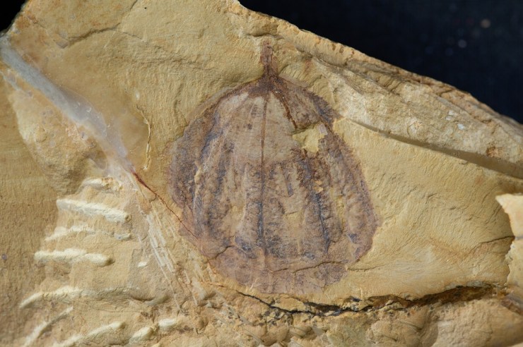
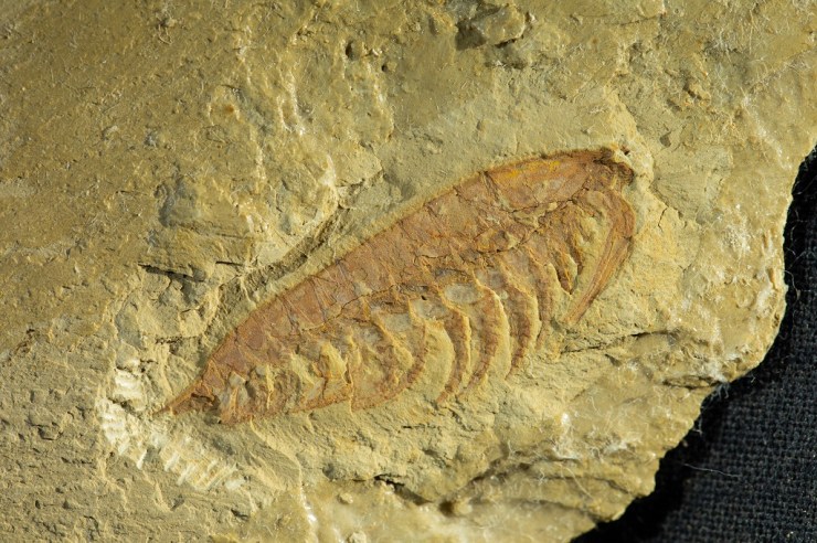
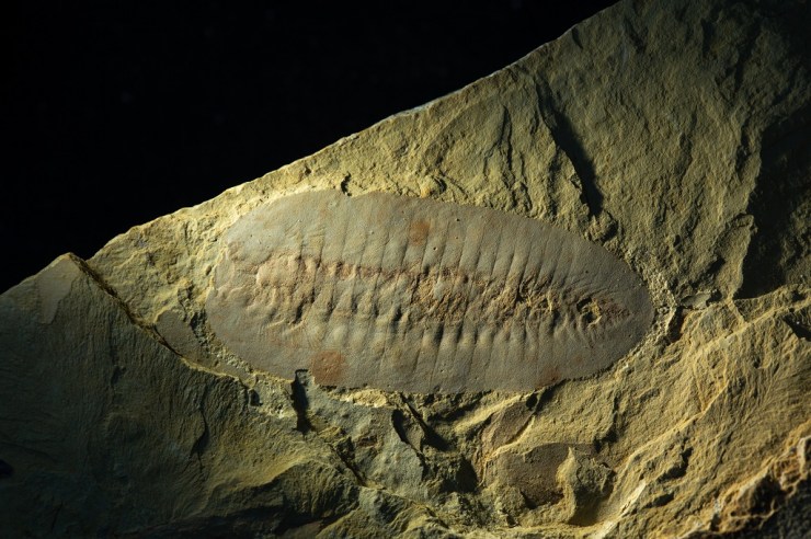
I quickly narrowed my interest down to fossils from the Cambrian period which are more complex life forms, more similar to life today. A collection of small fossils from the Chengjiang fossil site in Yunnan province, China was the inspiration for some initial observational drawings.

Abigail Harris - sketches for artwork showing reconstruction of Cambrian ocean animal life
A sketchbook page showing initial sketches and observations of Onychodictyon
Final illustration of Cotyledion

After returning to Plymouth University, I began to develop these initial sketches and observations, continuing to research the Chengjiang material and learning more about the characteristics of some of the creatures preserved as fossils.

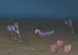
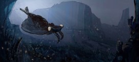
I wanted to create an under-the-sea ecology reconstruction showing a diversity of life forms, focusing on Onychodictyon, Cotyledion, Cricocosmia, Luolishania, and Paradiagoniella.

A five-step process was used for each reconstruction. Initially, I would sketch the fossil as I saw it, then I would research the characteristics and features of that animal, making a list of things to include in my drawing. A second drawing would then include all of these characteristics, not just what was initially visible in the fossil.

These rough sketches were then sent to the scientists for feedback, helping me to redraw and paint the illustrations with watercolour, before scanning and digitally editing each painting. Lastly, I created a background and added my illustrations.

Initial under under the sea ecology reconstruction.

Although the reconstructions were not completely finished by the time of my project deadline, I returned to the Museum in July and was given a tour of the First Animals exhibition by Deputy Head of Research Imran Rahman, as well as the opportunity to discuss how to improve my artworks for accuracy.

Another round of sketching and painting led to the final piece, shown at the start of this article, complete with an added digital background of the seafloor, and darkened to reflect the murky world of a Cambrian ocean, 50 metres below the surface.

Exceptional Chinese fossils come to Oxford in new partnership

by Imran Rahman, Deputy Head of Research

China is world-famous for its unique and exceptionally preserved fossils, which range from some of the oldest animals on Earth, to spectacular feathered dinosaurs. We are therefore very excited to announce that the Museum, along with other institutions from across Europe, is a partner in a major new venture with Yunnan University in China: the International Joint Laboratory for Palaeobiology and Palaeoenvironment.

Collaboration between this Museum and Yunnan University dates back to the 1990s, driven by the work of Professor Derek Siveter – a former Senior Research Fellow and current Honorary Research Associate at the Museum. He collaborated with Professor Hou Xianguang, director of the International Joint Laboratory for Palaeobiology and Palaeoenvironment, to study fossils from the internationally renowned Chengjiang biota, which was discovered by Hou Xianguang in 1984.

Museum researchers Duncan Murdock, Jack Matthews and Derek Siveter (l-r) visit the Precambrian-Cambrian Section

The Chengjiang fossil site is important and exciting because it preserves both the soft and hard parts of a range of early animals. This fossil record captures the rapid diversification of life about 520 million years old – in an event referred to as the Cambrian explosion. Derek Siveter was instrumental in a successful bid to have the Chengjiang biota designated a UNESCO World Heritage site in 2012, preserving it for future generations.

In December 2018, Museum researchers Duncan Murdock, Imran Rahman and Jack Matthews travelled with Derek to Kunming, China, for the first meeting of the International Joint Laboratory for Palaeobiology and Palaeoenvironment. The lucky researchers spent three days on field trips to the region’s most spectacular fossil sites, including Lufeng World Dinosaur Valley and the Chengjiang biota itself, followed by two full days of scientific talks and discussions.

The International Joint Laboratory is funded by the Ministry for Education of China and includes the University of Leicester, the Natural History Museum, London, the University of Munich, and the Bavarian State Collection of Zoology, along with Oxford University Museum of Natural History and Yunnan University.

The arthropod Haikoucaris ercaiensis. Sometimes referred to as a ‘short-great-appendage’ arthropod, Haikoucaris had a pair of prominent grasping appendages adjacent to the head (right-hand side of this image). Credit: Scott Billings
The arthropod Leanchoilia illecebrosa. Sometimes referred to as a ‘short-great-appendage’ arthropod, Leanchoilia illecebrosa had a pair of prominent grasping appendages adjacent to the head (right-hand side of this image). Credit: Scott Billings

A significant first outcome of this new partnership will be the loan of iconic Chengjiang fossil specimens from Kunming to Oxford for our First Animals exhibition which opens on 12 July and runs until February 2020. Most of these fossils have never been outside of China before, and some have never been seen by the public before. We invite you to visit First Animals to see these exceptional fossils first hand!

The arthropod Saperion glumaceum. Saperion had a flattened, segmented body and jointed appendages (not visible in this specimen). Credit: Scott Billings.

Top image: The comb jelly Galeactena hemispherica. Unlike modern comb jellies, which are soft-bodied animals, Galeactena and its relatives had hardened ‘spokes’ on the sides of the body (appearing as dark bands in this photograph). Credit: Scott Billings.

Lynn Margulis and the origins of multicellular life

To mark International Women’s Day Professor Judith Armitage, lead scientist on the Bacterial World exhibition, reflects on the ground-breaking – and controversial – work of evolutionary biologist Lynn Margulis

Iconoclastic, vivacious, intuitive, gregarious, insatiably and omnivorously curious, partisan, bighearted, fiercely protective of friends and family, mischievous, and a passionate advocate of the small and overlooked.

Lynn Margulis at the III Congress about Scientific Vulgarization in La Coruña, Spain, on November 9, 2005. Image: Jpedreira, CC BY-SA 2.5

These are all words used to describe evolutionary biologist and public intellectual Lynn Margulis. Intellectually precocious, Margulis got her first degree from the University of Chicago aged 19, but it was her exposure to an idea about the evolution of a certain type of cell that ignited a lifelong focus of her work.

This idea claimed that eukaryotic cells – cells with a nucleus, found in all plants and animals, but not bacteria – were first formed billions of years ago when one single-celled organism – a prokaryote – engulfed another to create a new type of cell. This theory, known as endosymbiosis, was laid down in a paper by Margulis in 1967. It brought her into conflict with others, including the so-called neo-Darwinists who believed in slow step-wise evolution driven by competition between organisms, not cooperation.

So what happened in the earliest evolution of these crucial cells? Initially, one bacterium ate a different, oxygen-using bacterium but didn’t digest it. Over time the two became interdependent and the bacterium took over almost all of the energy-generating processes of the host cell, becoming what we now call a mitochondrion. This allowed the cell to evolve into bigger cells and eventually form communities and develop into multicellular organisms.

Animal cells evolved when one single cell, possibly an archaeon, engulfed an aerobic bacterium – one that used oxygen to release energy. The bacterium evolved into the mitochondrion, the powerhouse of the cells of humans and other animals. A similar process created the chloroplasts found in plant cells.

These early mitochondria-containing organisms continued to eat other bacteria, and on more than one occasion they ate a photosynthesising cyanobacterium which evolved into a chloroplast, a structure now found inside plant cells.

The revolution in DNA sequencing that started in the 1970s, and continues today, eventually vindicated Margulis’ position on this ancient sequence of events. It revealed that chloroplasts and mitochondria both contain DNA with the same ancestry as cyanobacteria and proteobacteria respectively. In other words, both chloroplasts and mitochondria have evolved from ancient bacteria.

Margulis’ enthusiastic support for these ideas led her to think about the role of biology in the geology of Earth and some of its major changes, in particular the oxygenation of the atmosphere by cyanobacteria around 2.5 billion years ago. Mitochondria use oxygen, and so must have evolved from bacterial ancestors that arose after the cyanobacteria started to produce oxygen through photosynthesis.

Margulis met Gaia theorist James Lovelock soon after her seminal publication on endosymbiosis. At the time, Lovelock was looking at the composition of the atmosphere and factors causing change, including oxygen levels. He was starting to think of the Earth as a system – Gaia as it became known – where the planetary environment is regulated and kept stable by biological activity.

This meeting brought together two scientific outliers. Together they produced highly controversial articles on the “atmosphere as a biological contrivance”. Lovelock believed in concentrating on examining the systems as they are now, while Margulis brought deep time and evolutionary depth into the picture.

Margulis’ ideas were not always right, and she was enormously controversial in her time, but she made people think again. And in doing so she moved our understanding of things as apparently academically distant as the evolution of tiny cells billions of years ago to the stability of Earth’s environment today.

Top image: Euglena, a single cell eukaryotic. By Deuterostome [CC BY-SA 3.0]

Life’s big bang?

by Harriet Drage and Scott Billings

You may have heard of the Cambrian Explosion, an ‘event’, starting roughly 540 million years ago, when all the major animal groups suddenly appear in the fossil record, an apparent explosion of life and evolution.

But was there really an evolutionary explosion of all these animal groups, or is the lack of evidence from earlier periods due to some peculiarity of the fossilisation process? The debate has rumbled on for a number of years.

Now, a new study from our research team, the University of Oxford’s Department of Zoology, and the University of Lausanne, claims that the early Cambrian saw the origins and evolution of the largest and most important animal group on Earth – the euarthropods – in a paper which challenges two major pictures of animal evolution.

Euarthropoda contains the insects, crustaceans, spiders, trilobites, and a huge diversity of other forms alive and extinct. They comprise over 80 percent of all animal species on the planet and are key components of all of Earth’s ecosystems, making them the most important group since the dawn of animals over 500 million years ago.

Exceptionally preserved soft-bodied fossils of the Cambrian predator and stem-lineage euarthropod Anomalocaris canadensis from the Burgess Shale, Canada. Top left: Frontal appendage showing segmentation similar to modern-day euarthropods. Bottom right: Full body specimen showing one pair of frontal appendages (white arrows) and mouthparts consisting of plates with teeth (black arrow) on the head. Images: A. Daley.

A team based at the museum, and now at Lausanne, conducted the most comprehensive fossil analysis ever undertaken on early euarthropods, to try and establish whether these animals really did emerge in the early Cambrian period, or whether fossilisation just didn’t occur in any earlier periods.

In an article published today in the Proceedings of the National Academy of Sciences they show that, taken together, the total fossil record does show a gradual radiation of euarthropods during the early Cambrian, 540-500 million years ago, challenging other ideas that suggest either a rapid explosion of forms, or a much slower evolution that has not been preserved in the fossil record.

Each of the major types of fossil evidence has its limitation and they are incomplete in different ways, but when taken together they are mutually illuminating
Professor Allison Daley

Reconstruction of the Cambrian predator and stem-lineage euarthropod Anomalocaris canadensis, based on fossils from the Burgess Shale, Canada. Reconstruction by Natalia Patkiewicz.

By looking at a huge range of fossil material the researchers ruled out the possibility that Pre-Cambrian rocks older than around 541 million years would not have preserved early euarthropods. The only plausible explanation left is that the origins of this huge animal group didn’t evolve until about 540 million years ago, an estimate which also matches the most recent molecular dating.

The timing of the origin of Euarthropoda is very important as it affects how we view and interpret the evolution of the group and its effects on the planet. By working out which groups developed first we can trace the evolution of physical characteristics, such as limbs.

Exploring all the evidence like this allows us to make an informed estimate about the origins of key animal groups, leading to a better understanding of the evolution of early life on Earth.

Model of the Cambrian stem lineage euarthropod Peytoia, based on fossils from the Burgess Shale. Top left: Closeup of the mouth parts and frontal appendages. Bottom right: Overall view of the body. Model and image: E. Horn.

What is a tree of life?

A phylogeny? An evolutionary tree? A cladogram? We see the branching lines of these diagrams in many museum displays and science articles, but what do they tell us and why are they helpful?

Duncan Murdock, research fellow, explains. 

You are a fish.

Starfish, jellyfish and cuttlefish are not fish.

Actually, no, there’s no such thing as a fish. Let’s take a step back…

The Jackson 5 – the ultimate singing family tree?
Credit: Wikimedia Commons

It all comes down to common ancestry. All life is related, and we can think of it in terms of a family tree (or ‘phylogeny’): Jackie, Tito, Jermaine, Marlon and Michael were all Jacksons. United not only by a collective inability to control their feet, but also by common descent – they are all their parent’s children*.

By tracing further and further back in MJs family tree we could define ever larger groups united by common ancestors, first cousins (grandparents), second cousins (great-grandparents), all the way to every human, every mammal, every animal, and eventually all life – we are family (ok, that was Sister Sledge, but you get the point).

In the case of the tree of life, species are at the tips of branches and their common ancestors are where branches meet. A true biological group consists of a common ancestor and all its descendants, and we can use characteristics common between two species to imply common descent. Siblings look a lot like each other because they have inherited much of their appearance via common ancestry (i.e. their parents). In a similar way, two closely related species will share lots of inherited characteristics.

However, things are not quite that simple. Wings of bats, birds and insects are not inherited from a common ancestor but independently evolved for the same purpose, in this case flight. To complicate things further, as species evolve they may lose features inherited from their ancestors that other descendants retain. Snakes have lost their limbs, but still sit in the same group as lizards. These problems can be overcome by looking at many characteristics at once, using genetic information to test predicted relationships, and adding fossils to the tree to track change or loss through time (as in snakes).

Birds, insects and bats have all evolved wings for flight, but did not inherit this feature from a common ancestor. This is a good example of convergent evolution.

So, what about fish? ‘Fish’ is used to refer to pretty much anything that swims in water, but this lifestyle in animals like starfish (a relative of crinoids and sea urchins), jellyfish (a relative of corals) and cuttlefish (a relative of squid and octopus) evolved independently from more familiar fish like cod and carp. So, they’re not really ‘fish’ at all. With that in mind, how can we be fish? Well, the last common ancestor of, say, hagfish, salmon, shark and lungfish, is also the common ancestor of frogs, lizards, cats and us! All four-limbed animals with backbones descend from a fish-like ancestor. To complicate things further some have adapted to life back into the water and look much more like a ‘fish’ again, like dolphins, seals and the extinct ichthyosaur. Without a tree of life, we could not begin to unravel the evolutionary path that lead to all the diversity of life we see today.

The Blue Fin Tuna on display in the Museum is definitely a fish… right?!

You are closer to a chimp than a monkey, closer to a starfish than a snail, and closer to a mushroom than a tree. And, of course, there’s no such thing as a fish, but they still go well with chips.

*Joseph Jackson and Katherine Scruse had ten children, including the members of the Jackson 5, twenty-six grandchildren and several great-grandchildren.


The ancient mariner

Helen J. Bullard is a PhD candidate at the University of Wisconsin–Madison whose research aims to tell the historical and cultural stories of the horseshoe crab. After visiting the museum, and reading the story of our Natural History After-School Club member’s horseshoe crab fossil find, Helen offered to write a guest post for the blog about these amazing, ancient mariners…

You’re reading this, so I’m guessing you like museums. But have you ever heard of living fossils? Animals such as sharks and crocodiles are often referred to as ‘living fossils’ because they appear pretty unchanged from their ancient fossilized relatives. Of course, by definition, you can’t be both alive and a fossil. But fossils allow us to become primary eyewitnesses to ancient life; we can literally see what life used to look like, how cool is that? They can also dole out some pretty valuable advice, if we just choose to listen.

This summer during a visit to England, I spent some time at the Museum studying another so-called living fossil, the horseshoe ‘crab’. The horseshoe crab is not actually a crab, but is instead more closely related to spiders, scorpions and ticks. In fact, they are the closest living relatives of the extinct trilobites. But unlike their famous trilobite cousins, horseshoe crabs have survived all five of Earth’s major mass extinction events. Today, as a direct result of their ability to survive, the four remaining species of horseshoe crab play a vital role in global medical safety.

The Museum’s fossil specimen of Mesolimulus walchi, from the Upper Jurassic (163-145 million years ago), Solnhofen Germany, shows how little the form of the horseshoe crab has changed since

Not only do living horseshoe crabs look very similar to their early relations, they are also able to survive surprisingly severe injuries that often leave them missing body parts. Being able to see, through fossil evidence, how little their form has changed over time has helped to uncover the answer to this secret superpower. It lies in a very special life-saving trick that the crabs have kept for millions of years: a coagulating blood protein.

Horseshoe crabs on display in the Museum may provide food for thought for visitors

The blood of the horseshoe crab is able to clot quickly if bacteria are introduced, preventing infection, and saving the crab’s life. Since this discovery in the 1970s, this life-saving protein has been extracted from horseshoe crab blood and used in human medicine to test the safety of vaccines, medical laboratories, intravenous drugs, implants, and much, much more. The chances are that you owe a great deal of gratitude to the horseshoe crab.

But after all that surviving, horseshoe crabs, like many species, are now struggling for survival. They are losing their spawning grounds because of coastal development, industry, housing, marinas and coastal defense structures; they are collected and killed by the millions for bait, and bloodlet in their hundreds of thousands for medical use every year. It is likely that horseshoe crabs will not survive much longer.

But don’t despair. Museums are critical because they hold collections that can unlock knowledge about environmental change, and we can use that knowledge to protect life. Of course, horseshoe crabs are not alone in telling their stories through the fossils they leave – natural history museums are full of stories in stone, bones, pollen, and other traces. If you want to learn about and protect biodiversity, visit your local museum, or support organisations like Oxford’s Environmental Change Institute.

And to help the ancient horseshoe crab itself, join in with the efforts of the Ecological Research and Development Group – the crabs have saved us, so let’s return the favour.