Bursting into life

By Ricardo Pérez-de la Fuente, Museum Research Fellow

One of the earliest and toughest trials that all organisms face is birth. In egg-laying animals, the egg shell that has protected the embryo during its early development ultimately becomes a hard barrier between the animal and its life out in the world. The bursting of the egg is literally a threshold moment, and there are many ways to crack an egg…

Some animals break the egg membranes using dissolving chemicals; others physically, mechanically tear their way through the shells. Among the latter, a great diversity of animals use specialised devices called egg bursters. These vary greatly among the many arthropods and vertebrates that use them, but perhaps the most famous example is the ‘egg tooth’ that is present on the beak of newborn chicks.
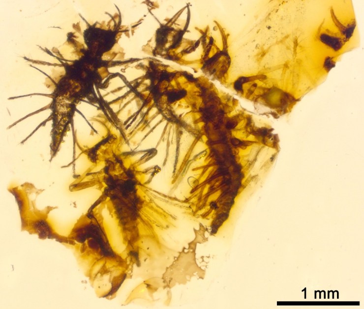
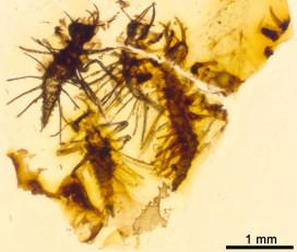
Four complete Tragychrysa ovoruptora newborns preserved together with egg shell remains and one egg burster. Modified from the open access Palaeontology paper.

My colleagues and I have found an exceptional fossil in 130 million-year-old Lebanese amber. Inside, trapped together are newborn larvae from Green Lacewings, the split egg shells from where they hatched, and the minute egg bursters that the hatchlings used to crack the egg. This is a first: no definitive evidence of these specialised egg-bursting structures had been reported from the fossil record of any egg-laying animals, until now.

The finding has been recently published as open access in the journal Palaeontology. Because multiple newborns were ensnared and entombed in the resin simultaneously, the fossil larvae have been described as the new species Tragichrysa ovoruptora, meaning ‘tragic green lacewing’ and ‘egg breaking’. A sad event, indeed, taking place in an ordinary day 130 million years ago in the Cretaceous forests of Lebanon, yet a happy circumstance now that we can take a privileged glimpse into the adaptations and behaviours of these fascinating tiny creatures.

The hatchlings from modern Green Lacewings open a slit on the egg with a ‘mask’ bearing a saw-like blade. Once used, this ‘mask’ is shed together with the embryonic cuticle and is left attached to the empty egg shell.

With the help of Amoret Spooner, Collections Manager at the Museum, egg clutches from modern green lacewings were found in the Museum collections. These eggs happened to have the intact egg bursters still attached to them, and proved to be crucial to understand that we had the same structures preserved in the amber together with the newborn larvae.

Two Tragychrysa ovoruptora newborns preserved together with egg shell remains and two visible egg bursters (right inset). Modified from the open access Palaeontology paper.

Green Lacewing larvae are small predators that often carry debris as camouflage, using their sickle-shaped jaws to pierce and suck the fluids of their prey. Whereas the larvae trapped in amber differ significantly from modern-day relatives, in that they possess long tubes instead of clubs or bumps for holding debris, the studied egg shells and egg bursters are remarkably similar to those of today’s green lacewings.

The larvae were almost certainly trapped by resin while clutching the eggs from which they had freshly emerged. Such behaviour is common among modern relatives while their body hardens and their predatory jaws become functional. Indeed, the two mouthparts forming the jaws are not assembled in most of the fossil larvae, which indicates, together with the large relative size of the head and legs, that they were recently born.
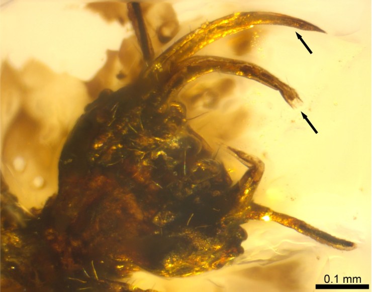
Detail of a head with the jaws still dislodged, indicating that the larva was recently hatched when it was ensnared by amber and the jaws had not yet had time to fully assemble.

It may seem reasonable to assume that traits controlling a life event as decisive as hatching would have remained largely unchanged during evolution. In fact, we see in very closely related insect groups different means of hatching that can entail the loss of the egg bursters. So the persistence of a hatching mechanism in a given animal lineage through deep time can’t be determined without direct proof from the fossil record.

Reconstruction of two Tragichrysa ovoruptora newborns clutching the eggs from where they recently hatched, moments before they were trapped by resin. Larvae colour and egg stalks are conjectural. Extracted from the open access Palaeontology paper.

This new discovery shows that the mechanism green lacewings use to crack the egg was already established 130 million years ago. Overall, it represents the first direct evidence of how insects hatched in deep time, egg-bursting their way through into life.

*

The hatching mechanism of 130-million-year-old insects: an association of neonates, egg shells and egg bursters in Lebanese amber by Ricardo Pérez-de la Fuente, Michael S. Engel, Dany Azar and Enrique Peñalver is published as open access in Palaeontology this month.

Imagining lost worlds

Earlier this year University of Plymouth illustration student Rachel Simpson teamed up with our research fellow Jack Matthews to ‘bring the oldest multi-cellular organisms back to life’. Rachel tells us about the process of working with some of the most ancient fossil material and reveals the results of her illustrations and modelling.

Illustration by Rachel Simpson, created in collaboration with the Museum

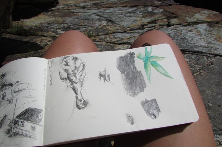
In August 2018 I was lucky enough to travel to Newfoundland, Canada with Dr Jack Matthews to learn about and illustrate some of the extraordinary fossils found there. A highlight of the trip was going down onto the fossil surface – known as the MUN surface – to look at examples of organisms such as Beothukis, Charnia and Primocandelabrum, all of which date from the Ediacaran period, over 550 million years ago.

The MUN surface is the location of the fossils that I had worked on for my university project. I had spent the previous months sketching, drawing and bringing these organisms back to life from silicon casts, so it was amazing to be able to see the real specimens in situ and to sketch from the fossil surface.

Sketching directly from the fossils also provided a new challenge as I was unable to control factors such as the lighting, which is crucial to seeing the fossils clearly. Nonetheless, I learnt a lot about drawing on location.

Sketching at the fossil surface

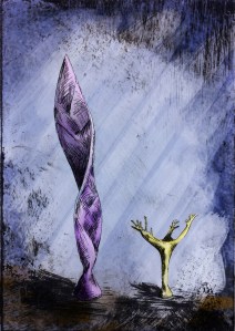
While visiting Port Union I was able to use some of the old printing presses held by the Sir William F. Coaker Heritage Foundation to create work inspired by the fossils I had seen in the surrounding area. I love using printmaking in my own illustrative practice so it was a great experience to get to use these old presses (image at top of article).

We also had the chance to give a radio interview and talk to the Port Union community about the work that Jack and I had done, showing how science and art can work together.

On my last day in Port Union I was invited by a local potter to make some ceramic representations of the fossils I had been drawing there. I created models of Fractofusus and Aspidella, and discovered that re-imagining something in three dimensions is a very different process to recreating it as a drawing.

Rachel created ceramic representations of some of the Ediacaran organisms

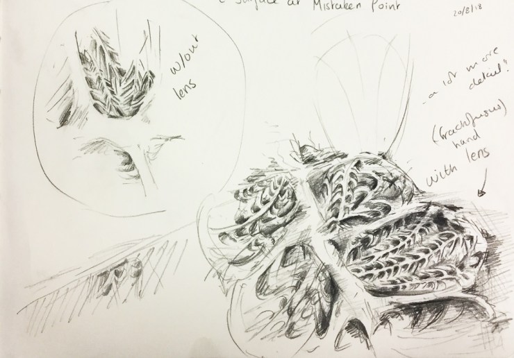
For the final three days of the trip we relocated from Port Union to Trepassey to visit the Mistaken Point UNESCO World Heritage Site. Here, I saw the highly preserved Fractofusus specimens and made some more sketches. Using a small hand lens I was able to draw all the details that are invisible to the naked eye.

Using a hand lens allowed Rachel to pick out details in the Fractofusus fossil

Drawing on location in Canada provided a better idea of the organisms in relation to other surrounding organisms, something that is more obscure when working from museum specimens. This definitely informed my practice and meant that artwork created after the trip was more representative of the science.

When I returned to England, I created some new prints inspired by my time in Newfoundland, the fossils that I saw, and the printing process I was able to use in Port Union.

A set of prints made by Rachel based on her work in Newfoundland

Crafty camouflage

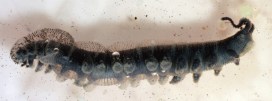
Last week we brought you snails that attach all manner of pebbles, fossils, corals and shark teeth to their shells. Today we give you a newly-discovered fossil green lacewing larva that attached pieces of soil to its body as an act of camouflage. Our research fellow Ricardo Pérez-de la Fuente, lead author of the new paper, explains…

Visual camouflage is one of the most successful survival strategies in nature. Camouflaging is usually defensive, allowing animals to be left unnoticed by their predators, but it can also be used aggressively by predators themselves to approach their prey undetected.

Some camouflaging animals can actively change their colouring to match that of the background ‒ a technique called crypsis. Others can make their bodies resemble elements of the environment, such as leaves or twigs, which is called mimicry.

Italochrysa italica, an extant green lacewing larva carrying a dense debris packet made of soil fragments. Taken from the open access publication Tauber & Winterton, 2014.

Yet another approach to camouflage involves collecting diverse materials from the environment and incorporating them on the animals’ bodies in order to better blend with the surroundings. This is known as debris-carrying, trash-carrying, or decoration, and it can be found across a wide variety of animals including sea urchins, gastropods, and arthropods, such as decorating crabs, or sand- and mud-covering spiders.

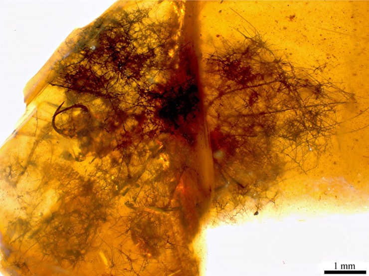
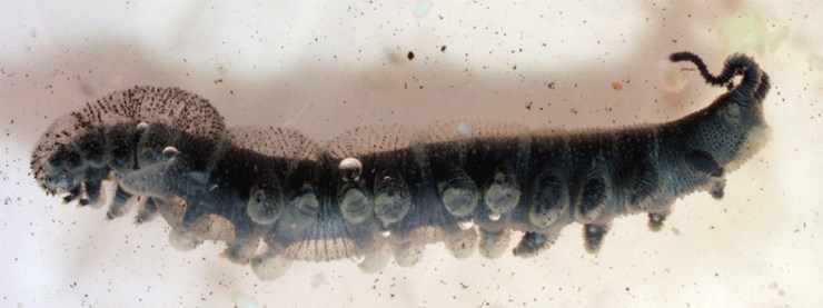
My colleagues and I have just published the discovery of a fossil green lacewing larva, pictured at the top of the article, that has been preserved carrying bits of soil that it used for camouflage and physical protection. It’s a new larval species just 1.5 mm in length, and is preserved in Early Cretaceous Lebanese amber. We have named it Tyruschrysa melqart after the Phoenician city of Tyre and its tutelary god Milk-Qart (if you want to learn the reasons behind this name check out our open access paper!).

Interpretative drawing of Tyruschrysa melqart: body in grey, ‘tubes’ with setae coloured according to which body part they are attached to, and soil debris in brown.

Green lacewing larvae are active predators that eat other insects such as aphids, using sickle-shaped ‘jaws’ to pierce their prey, suck out their fluids and liquefy their tissues; eating is easier when there is no need to chew! Some green lacewing larvae are debris carriers, entangling all kinds of debris among their velcro-like ‘hairs’ called setae, which extend from relatively short ‘bumps’ on their backs. This debris is carefully selected and gathered with meticulous head and body movements to form a so-called debris packet on the back of the insect.

‘Tubes’ bearing setae of Tyruschrysa melqart, with detail of their mushroom-shaped endings (bottom), used for anchoring bits of soil.

The new fossil and similar ones described from younger Cretaceous ambers differ from modern relatives because instead of short ‘bumps’ with setae on their backs they have relatively long ‘tubes’, giving them a bizarre appearance.

These tubes have setae with mushroom-shaped endings of a kind never seen before in extinct or living green lacewing larva species. The mushroom-shaped ending is a special adaptation to anchor debris, which in the case of Tyruschrysa melqart are fragments of soil.

Hallucinochrysa diogenesi, another Cretaceous green lacewing larva bearing long ‘tubes’ with setae on its back, but carrying a debris packet made of plant hairs (trichomes). Preserved in Spanish amber (105 million years old).

It was already known that Cretaceous green lacewing larvae like Tyruschrysa had long tubes on their backs and that they collected plant hairs and other plant material to construct their packet of debris. But thanks to the new discovery we now know that these immature insects also used bits of soil, and that in the deep past debris packets were probably as diverse as those we see today.

Green lacewing larvae have been gathering debris to camouflage and protect themselves for about 130 million years, giving rise to the different body adaptations we see amongst these fascinating tiny collectors.

‘A soil-carrying lacewing larva in Early Cretaceous Lebanese amber’ Ricardo Pérez-de la Fuente, Enrique Peñalver, Dany Azar and Michael S. Engel is published as open access in Scientific Reports this month.

Death, decay and fossilization

By Duncan Murdock, Research Fellow

Our oceans, rivers and lakes teem with life of all shapes and sizes, and have done so for hundreds of millions of years. We can get a glimpse of the wonderful diversity of life deep in the Earth’s past from fossils that can be found in the rocks beneath our feet. But the fossil record is as much a history of death as it is of life.

All animals die, in huge numbers every day, but the sea beds and forest floors of the Earth are not filling up with their remains. Decay is as inevitable as death. This is good news for those left behind, but bad news for fossil hunters.

Being preserved as a fossil is very much the exception, not the rule, and the chances of anything surviving the various processes by which the component parts of an animal are lost forever are vanishingly small, even for hard parts like shells, teeth and bones. For the ‘soft’ parts of animals, such as the muscles, eyes, guts and nerves, it is nearly impossible.

But ‘nearly impossible’ is good enough when you can consider every animal that ever lived, or more importantly, died. In exceptional circumstances ‘soft’ tissues do become fossils, and when they do they invariably give an unrivalled view of an otherwise completely lost world.

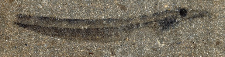
Although exceptionally well preserved, this fossil of a jawless fish is not entirely complete. Some features have been preserved, like the prominent dark eye spot and gill supports just beneath, but others, such as the guts and fins, have rotted away before they could be preserved. Image: Mark Purnell, Sarah Gabbott, Robert Sansom (University of Leicester)

We know from these exceptional fossils that the path from death to fossil is not random. Yes, you have to be lucky, but the odds are very much stacked towards certain combinations of who, what, where and when.

Furthermore, decay is not the whole story. Not only does anatomical information have to survive decay, it has to undergo parallel (but distinct) processes of preservation – conversion into materials that are stable over millions of years as part of a rock. It is the balance between the loss and retention of information that seals the fate of an organism’s remains.

Three hundred million years ago, a small worm gulped its last breath and died. Its body began to rot and, were it not for the peculiar conditions of the sediments it was laid to rest in, would have been lost forever. Fortunately for us, what remained was preserved in rock – a rotten fossil. But how much rotted away before it was fossilized? By decaying modern relatives in the lab we can model this missing history, and build better-informed reconstructions of extinct animals. Image: Duncan Murdock

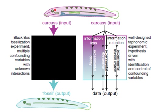
Left with only the lucky few, the parts of animals where retention exceeded loss, the fossil record is profoundly biased. One way to unravel this lost history of loss is it to conduct experiments, replicating decay and preservation. However, trying to make fossils in the lab, by contriving one particular set of conditions, is fiendishly complex – there are simply too many variables to set.

I have been working with researchers from the Universities of Leicester, Bristol, Manchester and University College Cork, and together we have described an alternative approach to unpack the ‘black box’ of fossilization and take each variable in turn, individually examining the different processes that result in retaining information as potential ‘fossils’ and, crucially, those that result in loss.

This cartoon illustrates the difference between experiments that attempt to replicate fossilization, treating the process as a black box, and the approach we are taking. The black box approach reveals little about the processes of information loss and information retention, the cumulative effects and interactions which ultimately results in a fossil (or, more often, not). Image: Purnell et al. 2018.

Ultimately this approach will allow more and more complex experiments to be designed, to unpick the interactions between the who, what, where and when in the lost history of death.

The techniques described here are published in Palaeontology today as ‘Experimental Analysis of Soft-Tissue Fossilization: Opening the Black Box‘, Purnell et al. 2018.

Bound by blood

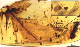
It may sound like we’ve stumbled into a script-writing session for Jurassic Park, but one of our research fellows, Dr Ricardo Pérez-de la Fuente, along with an international team, has discovered a parasite trapped in amber, clutching the feather of a dinosaur. This small fossilised tick, along with a few other specimens, is the first direct evidence that ticks sucked the blood of feathered dinosaurs 100 million years ago. Ricardo tells us all about it…

The paper that my colleagues and I have just published provides evidence that ticks fed from feathered dinosaurs about 100 million years ago, during the mid-Cretaceous period. It is based on evidence from amber fossils, including that of a hard tick grasping a dinosaur feather preserved in 99 million-year-old Burmese amber.

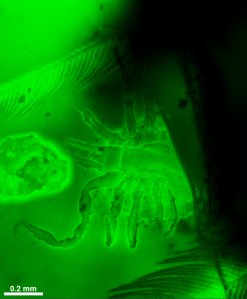
Fluorescence detail of the studied hard tick grasping a dinosaur feather. Extracted from the publication.

The probability of the tick and feather becoming so tightly associated and co-preserved in resin by chance is virtually zero, which means the discovery is the first direct evidence of a parasite-host relationship between ticks and feathered dinosaurs.

Fossils of parasitic, blood-feeding creatures directly associated with remains of their host are exceedingly scarce, and this new specimen is the oldest known to date. The tick is an immature specimen of Cornupalpatum burmanicum; look closely under the microscope and you can see tiny teeth in the mouthparts that are used to create a hole and fix to the host’s skin to suck its blood.

The structure of the feather inside the amber is similar to modern-day bird feathers, but it could not belong to a modern bird because, according to current evidence at least, they did not appear until 26 million years later than the age of the amber.

Feathers with the same characteristics were already present in multiple forms of theropod dinosaurs –  the lineage of dinosaurs leading to modern birds – from ground-runners without flying ability, to bird-like forms capable of powered flight. Unfortunately, this means it is not possible to determine exactly which kind of feathered dinosaur the amber feather belonged to.

But there is more evidence of the dinosaur-tick relationship in the scientific paper. We also describe a new group of extinct ticks, created from a species we have named Deinocroton draculi, or “Dracula’s terrible tick”. These novel ticks, in the family Deinocrotonidae, are distinguished from other ticks by the structure of their body surface, palps and legs, and the position of their head, among other characteristics.

Blood-engorged Deinocroton draculi tick (female). Extracted from the publication.

This new species was also found sealed inside Burmese amber, with one specimen remarkably engorged with blood, increasing its volume approximately eight times over non-engorged forms. Despite this, it has not been possible to directly determine its host animal:

Assessing the composition of the blood meal inside the bloated tick is not feasible because, unfortunately, the tick did not become fully immersed in resin and so its contents were altered by mineral deposition.
Dr Xavier Delclòs, an author of the study from the University of Barcelona and IRBio.

But there was indirect evidence of the likely host for these novel ticks in the form of hair-like structures called setae from the larvae of skin beetles, or dermestids, found attached to two Deinocroton ticks preserved together. Today, skin beetles feed in nests, consuming feathers, skin and hair from the nest’s occupants. But as no mammal hairs have yet been found in Cretaceous amber, the presence of skin beetle setae on the two Deinocroton draculi ticks suggests that their host was in fact a feathered dinosaur.

The hair-like structures, or setae, from skin beetles (dermestids) found attached to two Deinocroton ticks fossilised inside amber, in comparison with extant ones. Modified from the publication.

Together, these findings tell us a fascinating story about ancient tick behaviour. They reveal some of the ecological interactions taking place among early ticks and birds, showing that their parasite-host relationship has lasted for at least 99 million years: an enduring connection, bound by blood.

The paper “Ticks parasitised feathered dinosaurs as revealed by Cretaceous amber assemblages” is published as open access in Nature Communications. Direct link: http://dx.doi.org/10.1038/s41467-017-01550-z

The ancient mariner

Helen J. Bullard is a PhD candidate at the University of Wisconsin–Madison whose research aims to tell the historical and cultural stories of the horseshoe crab. After visiting the museum, and reading the story of our Natural History After-School Club member’s horseshoe crab fossil find, Helen offered to write a guest post for the blog about these amazing, ancient mariners…

You’re reading this, so I’m guessing you like museums. But have you ever heard of living fossils? Animals such as sharks and crocodiles are often referred to as ‘living fossils’ because they appear pretty unchanged from their ancient fossilized relatives. Of course, by definition, you can’t be both alive and a fossil. But fossils allow us to become primary eyewitnesses to ancient life; we can literally see what life used to look like, how cool is that? They can also dole out some pretty valuable advice, if we just choose to listen.

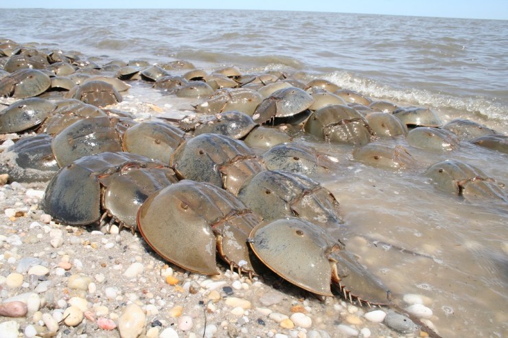
This summer during a visit to England, I spent some time at the Museum studying another so-called living fossil, the horseshoe ‘crab’. The horseshoe crab is not actually a crab, but is instead more closely related to spiders, scorpions and ticks. In fact, they are the closest living relatives of the extinct trilobites. But unlike their famous trilobite cousins, horseshoe crabs have survived all five of Earth’s major mass extinction events. Today, as a direct result of their ability to survive, the four remaining species of horseshoe crab play a vital role in global medical safety.

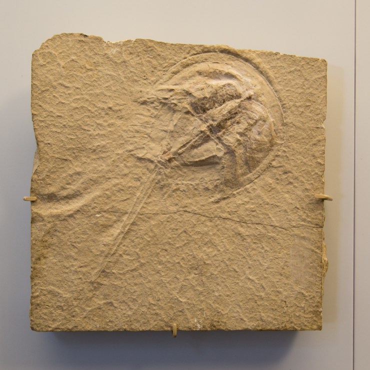
The Museum’s fossil specimen of Mesolimulus walchi, from the Upper Jurassic (163-145 million years ago), Solnhofen Germany, shows how little the form of the horseshoe crab has changed since

Not only do living horseshoe crabs look very similar to their early relations, they are also able to survive surprisingly severe injuries that often leave them missing body parts. Being able to see, through fossil evidence, how little their form has changed over time has helped to uncover the answer to this secret superpower. It lies in a very special life-saving trick that the crabs have kept for millions of years: a coagulating blood protein.

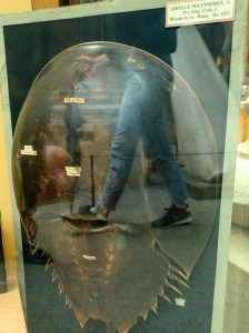
Horseshoe crabs on display in the Museum may provide food for thought for visitors

The blood of the horseshoe crab is able to clot quickly if bacteria are introduced, preventing infection, and saving the crab’s life. Since this discovery in the 1970s, this life-saving protein has been extracted from horseshoe crab blood and used in human medicine to test the safety of vaccines, medical laboratories, intravenous drugs, implants, and much, much more. The chances are that you owe a great deal of gratitude to the horseshoe crab.

But after all that surviving, horseshoe crabs, like many species, are now struggling for survival. They are losing their spawning grounds because of coastal development, industry, housing, marinas and coastal defense structures; they are collected and killed by the millions for bait, and bloodlet in their hundreds of thousands for medical use every year. It is likely that horseshoe crabs will not survive much longer.

But don’t despair. Museums are critical because they hold collections that can unlock knowledge about environmental change, and we can use that knowledge to protect life. Of course, horseshoe crabs are not alone in telling their stories through the fossils they leave – natural history museums are full of stories in stone, bones, pollen, and other traces. If you want to learn about and protect biodiversity, visit your local museum, or support organisations like Oxford’s Environmental Change Institute.

And to help the ancient horseshoe crab itself, join in with the efforts of the Ecological Research and Development Group – the crabs have saved us, so let’s return the favour.