Tales of Iguanodon Tails
By Leonie Biggenden, Volunteer
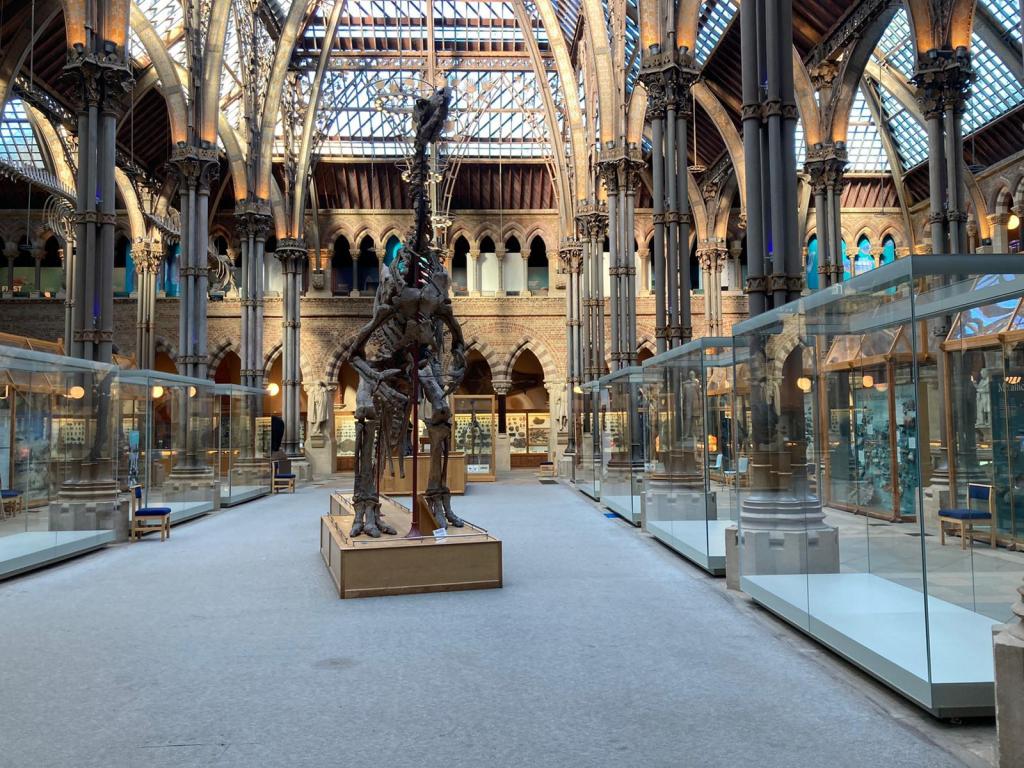
As one of our many invaluable volunteers, Leonie Biggenden has regularly helped to run our Science Saturdays and Family Friendly Sunday activities, both of which take place under the watchful eyes of the large T. rex and Iguanodon skeletons in the Museum’s main court. Having spent so much time beside the Iguanodon, and with a lack of in-person volunteering opportunities in recent months, Leonie decided to find out some of the history of this striking cast. For Volunteers Week this week, she shares what she discovered…
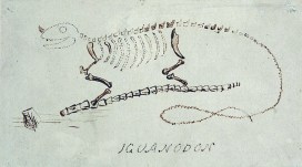
Next year will be the 200th anniversary of the discovery, by a roadside in Sussex, of the first Iguanodon teeth. Found by Mary Mantell in 1822, her husband Gideon saw their similarity with the teeth of modern iguanas and suggested they were from a huge, ancient, herbivorous lizard. He called the animal Iguanodon, and you can see his sketch reconstruction at the top of this post.
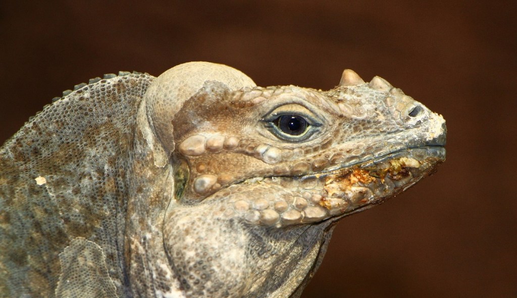
However, as an amateur palaeontologist, Gideon Mantell was not initially taken seriously by the scientific establishment. Some claimed the teeth were actually from a rhinoceros, or even a pufferfish! But in 1834, more complete remains were found by workmen who had accidentally blown up a slab of rock in a quarry near Maidstone, Kent. Iguanodon became a rock star of the dinosaur world, being only the second dinosaur – and the first herbivorous one – to be named (the first was the carnivorous Megalosaurus – another famous Museum specimen).
Twenty years later, a model of an Iguanodon was constructed by sculptor Benjamin Waterhouse Hawkins as one of a set of 30 life-sized models of extinct animals for the relocated Crystal Palace Gardens in South London. It was mounted in a rhinoceros-like pose, with what we now know as a thumb spike placed as a nose horn. Scientists always look to the information they have available to them, including observation of living animals, and there is an iguana called Cyclura cornuta – the Rhinoceros Iguana – which does indeed have nose horns, so at the time the nose horn made sense.
Another 20 years on and a most significant find was made in southern Belgium. In February 1878, more than 30 fully articulated, adult Iguanodon fossil skeletons were found by miners Jules Créteur and Alphonse Blanchard, 322 m deep in the Sainte Barbe coal mine. Louis de Pauw from the Belgian Royal Museum of Natural History started to excavate the skeletons. It was a risky undertaking. In August an earthquake cut them off for two hours, and in October they were forced to return to the surface as the mine flooded.
The fossils were wrapped in damp paper, covered in protective plaster, and divided into 600 blocks. Each specimen was given a number and each block a letter, to record their exact positions in the mine. The 130 tonnes of specimens, rock, iron reinforcing rods, and plaster were then brought to the surface of the mine by horse drawn trucks and transported to Brussels.
For the first time, scientists, and later the public, could see complete dinosaur skeletons. This was important because scientists learned that the unusual spike found in the scattered fossils in the UK was a thumb spike rather than a nose horn, and they ditched rhino resemblance too, though not in time for the Crystal Palace reconstruction!
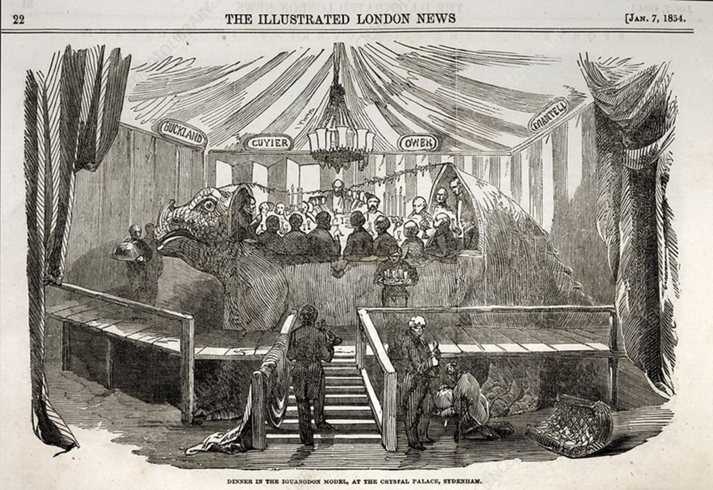
Illustrated London News January 7 1854, page 22. The Iguanodon model at the Crystal Palace in London was large enough for several people to dine inside it. 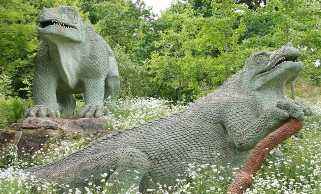
The models now in Crystal Palace park in South London. In these reconstructions the thumb spike was placed as a nose horn, and the animal is positioned in a rhinoceros-like pose.
Image: Chris Sampson, CC BY 2.0 , via Wikimedia Commons
In 1882, de Pauw began assembling at least 38 Iguanodon skeletons under instruction from Louis Dollo, another famous Belgian palaeontologist. The aim was to put them in their most probable living position. A room with a high ceiling was needed because of their size, and a chapel was chosen. Scaffolding was built with hanging ropes being adjusted so the fossilized bones could be moved into their most likely position and then fixed and reinforced with iron rods.
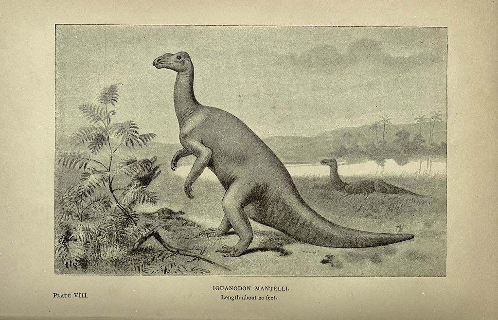
Early reconstructions of Iguanodon showed the dinosaur standing in a kangaroo-like stance. Image: Hutchinson, H. N., Public domain, via Wikimedia Commons 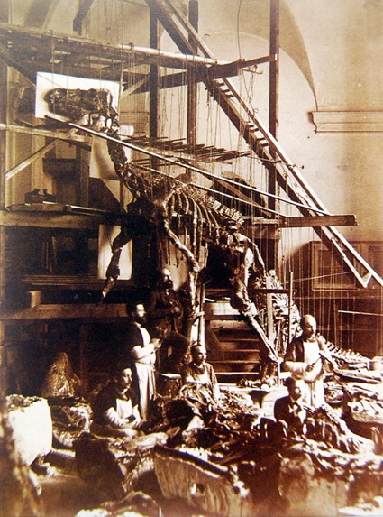
Workmen mounting the first Iguanodon bernissartensis skeleton in the St. George Chapel in Brussels, 1882.
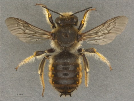
Iguanodon bernissartensis, like the one on display here in the Museum, was a new species, named in 1881. It lived about 125 million years ago. The first assembly was revealed in 1882 and went on public display in Brussels in 1883. Points of reference used for the pose were the skeleton of a cassowary and a kangaroo.
On the Museum’s cast skeleton you can see rod-like structures going across the blade-like, bony processes on the back. These are ossified, or hardened, tendons and would help to stiffen the tail and therefore restrict its movement. They have been broken where the bend in the tail was made to resemble a kangaroo-like stance. The displacement shows that the true position of the tail should be straight.
But having such a straight tail would mean that the Iguanodon would need its head and arms nearer the ground for better balance. The strong hind limbs suggest it would usually walk on two legs with its tail held aloft, as does the fact that fossil Iguanodon footprints are three-toed, and the three-toed limbs are the back ones.
By the end of 1883, six Iguanodons had been mounted this way and positioned in their own glass cage in the courtyard of the Brussels museum. So Iguanodon was one of the very first dinosaurs to be recovered in its entirety and mounted in three dimensions as though a living animal!
—
Leonie is a longstanding Public Engagement volunteer at the Museum. Unable to volunteer in the normal way during the lockdown, she researched the history of this favourite specimen and shared what she learned in a talk for other volunteers as part of an online ‘social’. This article has been adapted from that presentation.