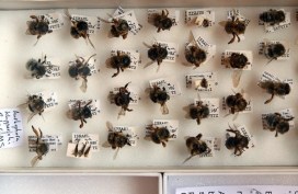
Plight of the bumblebee

nhm16

The ecological importance of bumblebees has become more widely appreciated in recent years, thanks to environmental campaigners and reports of species decline, and even some extinctions, in the UK.

To look at this issue, we have recently teamed up again with arts-science organisation Pale Blue Dot, which is launching a new research project to investigate why some species of bumblebee are declining and to raise awareness about the ultimate impact this has on people.

Here, Pale Blue Dot co-founder Jane King explains how the Bees & Weeds project brings together art students, public engagement, the Museum’s collections and a leading bumblebee scientist…

*

On 9 September we launched our latest project – Bees & Weeds – with the Museum of Natural History, building on our previous collaboration for the Lost & Found exhibition. We were joined by over 50 art students from Banbury & Bicester College to highlight the plight of the bumblebee, revealing how its decline is impacting everything from what we eat to where we live and work.

Amoret Spooner displays drawers from the collections

The students spent some time looking at methods of insect labelling and notation, before heading behind the scenes with entomologist Amoret Spooner to the Huxley Room, the location of the Great Debate on Darwin’s Theory of Evolution, which took place in 1860.

Amoret provided an insight into taxonomy – the science of species classification – as well as her work on the conservation of specimens. We visited the huge archive of bee specimens and learnt about some of the research that scientists are currently carrying out on UK bumblebee species to help prevent further decline.
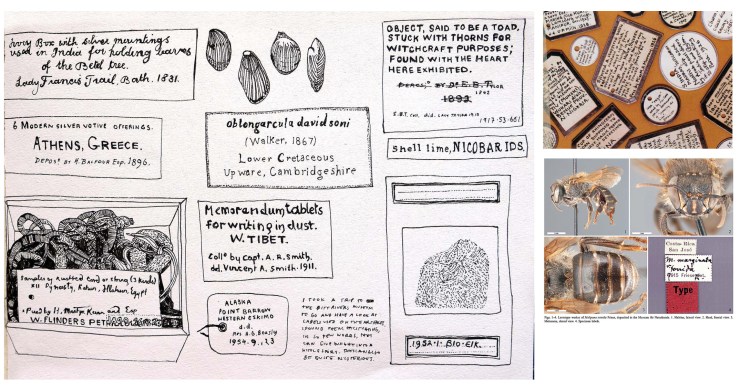
Student sketches of labelling and notation

During spring 2015, the art students from the Banbury & Bicester College, as well as students from Oxford Brookes University, will make and install hundreds of cycle seat covers on bikes in and around the Oxford city. The seat covers will carry messages about bumblebee decline in the UK countryside, showing how much we depend on their pollination services, which far outstrip those of the honeybee in their value to UK food production.

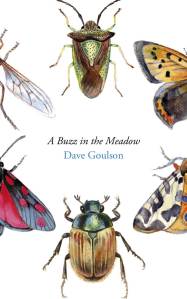
We are also working with Professor Dave Goulson from the University of Sussex, one of the world’s most important bumblebee scientists. He will present his research showing how well bumblebees are doing in gardens compared to the countryside, as well as the optimum range of flowering plants needed to help them thrive. Dave’s book, A Sting in the Tale, is already a best-seller, and the sequel – A Buzz in the Meadow – was published on 4 September.

Dave will also be speaking about his new book at the Museum on Thursday 9 October at 7pm. Book your tickets for that via Waterstones here.

Artwork from the Bees & Weeds project, together with cycle seat covers and bike paraphernalia, will be on show and for sale in the Old Fire Station in Oxford from next spring. If you cycle in Oxford, you may be lucky enough to receive one!

Pale Blue Dot is an arts-science organisation helping scientists to communicate their research to the public. It promotes an interdisciplinary approach to learning, living and working through exhibitions, publications and happenings.

Jane King – co-founder, Pale Blue Dot
Heads up… what’s on


CraspedophoraKeulemans crop

We try hard not to use this blog simply as a promotional tool for the Museum’s events, but thought that when a new quarterly programme is published you might like to know so you can have a look and pick out anything you fancy.

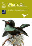
Oct-Dec prog coverSo this is just a short post to let you know that the October-December programme is now out. Highlights include a headline runway show for Oxford Fashion Week, on Saturday 8 November, with designs inspired by the beauty and variety of Birds of Paradise.

We have some Arctic-themed activity later in November, with the Northern Lights late night event for Christmas Light Festival on Friday 21 November, and a special talk by Museum director Professor Paul Smith, The Arctic – A Natural History, on Tuesday 18 November. Free tickets are available now.

Attachment-1Finally, there was a late arrival that didn’t quite make it into the programme leaflet, but is not to be missed: Dave Goulson, founder of the Bumblebee Conservation Trust, will talk about his latest work, A Buzz in the Meadow, on Thursday 9 October. Tickets can be booked here, and a limited number of early bird tickets include a behind-the-scenes tour in our entomology department.

With plenty of family events too, hopefully there’s something for everyone in the programme. If you’d like to be added to our general mailing list just drop me a line at communications@oum.ox.ac.uk and I’ll add your email address.

Scott Billings – Public engagement officer

Ready for your close-up?

IMG_3684
Image copyright Richard Kelsall

They say a picture is worth a thousand words, and at the Museum we make thousands of pictures: pictures to document, pictures to investigate, and pictures to wow. We use a lot of different imaging techniques too, from standard close-up photography to scanning electron microscopy, which reveals the most minute details.

To coincide with the final week of the Wildlife Photographer of the Year exhibition here, on Saturday 20 September we held a new adult workshop to give people some hands-on experience of some of these processes. Imaging Techniques in Modern Natural History gave participants the chance to get up close to some wonderful specimens and make their own images to take home.

I had planned to review the day here, but Rose Parkin, who took part in the workshops, very helpfully sent in her own write-up of the sessions. So here’s a special guest post from Rose, along with some pictures taken by people on the day.

*

By Rose Parkin

When I signed up for the digital imaging course I expected a fairly dry, tech-heavy day. Instead, the experience was really exciting. Not only did it provide hands-on experience of viewing and recording images with new technology, it also gave me a brief glimpse behind the scenes of my favourite museum.

Laser Scanning and Digital Modelling
For our first session our small group was led through a maze of corridors by Sarah Joomun, the Documentation Officer, to the laser scanning lab. It sounded a bit futuristic, and it turned out that it looks that way too. Sarah popped a fossil onto a mount, clicked a few buttons and red lasers appeared, scanning the fossil’s surface while it rotated. After ten minutes the first 3D image of the fossil was produced – a beautiful net of triangles, which looked like a teleporting object in a science fiction film.

Laser scanning Image copyright: Tom Nicholson-Lailey
Laser scanning
Image copyright Tom Nicholson-Lailey

Sarah turned the fossil and scanned it again. The challenge was then to fit  these two images together to make a complete 3D model. Amazingly, this technique enables other palaeontologists around the world to see and replicate, with the use of a 3D printer, the exact size and shape of a fossil without it ever leaving the museum.

Multi-plane Microscope Photography
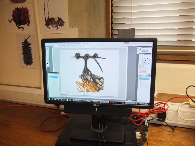
Our next session was upstairs, with artist-in-residence and photographer Katherine Child. Even though we were close to the main corridor of the museum it felt like a real working space, crammed full of equipment and insect specimens. Katherine had chosen the tiniest of insects for us to photograph with the multi-plane microscope. It looked like a small seed with some barely visible limb-like protrusions.

Multiplane photography. Image copyright Rose Parkin
Multiplane photography.
Image copyright Rose Parkin

But under the microscope a wonderfully strange insect became visible, with the most bizarre appendages and bright orange legs. While the microscope already showed a great deal of detail the multi-plane photography captured an incredibly crisp image. The microscope takes large numbers of photos of the specimen, using different focal planes each time, then the focussed elements are all stacked together to produce a crystal clear photograph.

Once we’d chosen and photographed some other insects from the collection and poked around the room a bit (finding a disturbing collection of large pickled spiders), we were taken on a tour of the entomology department. Katherine led us through corridors of offices and labs, up to a stunning store room that felt almost church-like, with rows and rows of cabinets full of fascinating insects.

Scanning Electron Microscopy
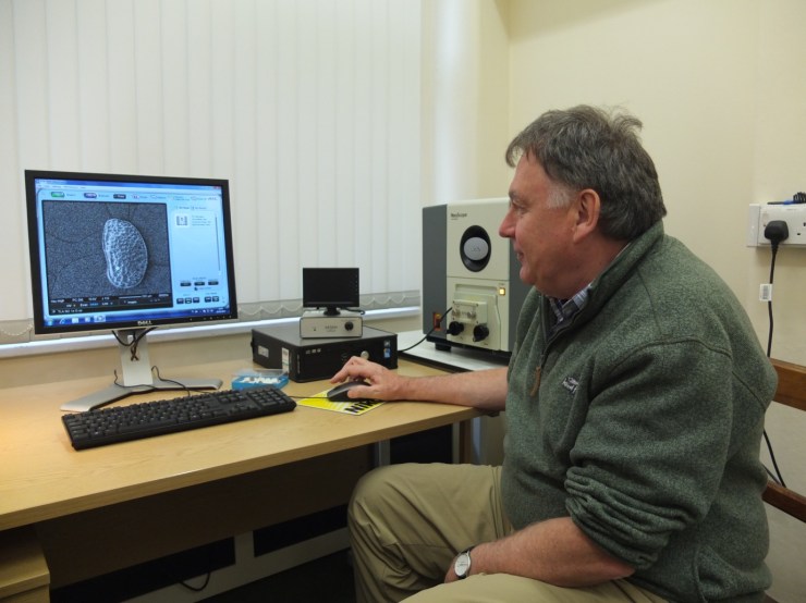
After lunch we had a laboratory session with museum director Paul Smith to look at sand under an electron microscope. Luckily, that was much more exciting than it sounds! The sand was taken from Dog’s Bay on the west coast of Ireland and is rich with a wide range of tiny fossilized organisms. Paul showed us how to carefully select individual microfossils from a tray using just a microscope and a paint brush.

PS and SEM
Professor Paul Smith demonstrates the scanning electron microscope. Image copyright Rose Parkin

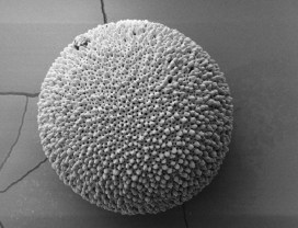
We then viewed some of the microfossils using a scanning electron microscope. This allowed us to see an incredible level of detail. The microscope was so powerful that we could see hair holes in a fossil the size of a grain of sand.

DSLR Macrophotography
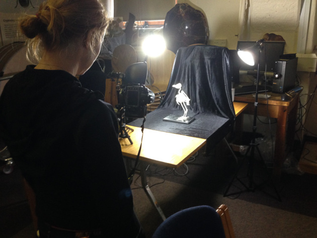
My final session was a crash course in macrophotography. Held in the seminar room, the low lighting and floor-to-ceiling collection of specimens lent an almost eerie feeling to the set-up.

Macrophotography. Image copyright Keith Barnes
Macrophotography.
Image copyright Keith Barnes
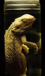
Bearded dragon. Image copyright: Rose Parkin
Bearded dragon.
Image copyright: Rose Parkin

Once prepped, we were let loose on four separate camera setups. Being able to choose and shoot at our own pace made this feel like a really creative experience. The help given by professional photographer Keith Barnes and public engagement officer Scott Billings was perfect – very hands on but not patronizing (despite my lack of DSLR experience).

With this digital imaging course the museum has created a really exciting snapshot of the work that goes on behind the scenes, reinforcing the fact that this impressive place is much more than just an ordinary museum.

A lesson from the past

PETM Foram

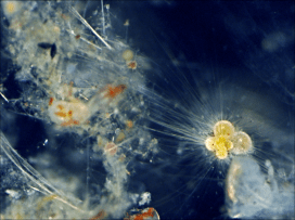
If you’re a regular reader of this blog you might have heard of Dr Tracy Aze already, and may even recognise the strangely-shaped specimen above as an example of planktonic foraminifera, the single-celled marine organisms that Tracy has been researching. This morning we have issued a press release about Tracy’s research which offers a warning from history about carbon emissions and global warming.

Surprisingly enough, the study shows how the fossils of these creatures hold clues to the impact on our oceans of man-made global warming. Around 56 million years ago, in a period known as the Palaeocene-Eocene Thermal Maximum (PETM), a rapid rise in greenhouse gases caused sea surface temperatures to rise as high as 40°C, with significant impacts on marine life.

Worryingly, the PETM – which lasted for around 170,000 years – saw the release of roughly the same volume of CO2 as expected from modern fossil fuel consumption. Tracy explains:

The amount of CO2 that is predicted to be released from the Industrial Revolution to around 100 years from now is roughly equivalent to what happened in the PETM. But the big difference is the rate of release: today we are releasing greenhouse gases at a far faster rate than 56 million years ago.

Although the research was conducted by Tracy, the project was led by Professor Paul Pearson of Cardiff University and funded by the UK Ocean Acidification research programme.

Tracy and her team used newly-extracted planktonic foraminifera fossils from Tanzania, dating from the PETM period. The tiny shells of these organisms contain different proportions of oxygen isotopes and these proportions are largely determined by the sea temperatures at the time. So the fossil shells offer a glimpse of the way sea temperatures were rising alongside the release of greenhouse gases, as well as a record of the relative abundance of this planktonic life in the oceans.

The PETM shows us that rapid increases in CO2 in the atmosphere have significant impacts on global temperatures, with the new information from our study site showing that tropical sea surface temperatures may have exceeded 40°C with an associated local disappearance of marine life.

The research paper, Extreme warming of tropical waters during the Paleocene–Eocene Thermal Maximum, was published in the September issue of Geology and is available as open-access.

Scott Billings – Public engagement officer


Smaller than a dodo

Grace Manley, one of our interns, writes about her work on planktonic foraminifera…

morethananintern's avatarMore Than An Intern

The museum not only contains a huge array of specimens both on display and on the collections, but is home to active research undertaken by research fellows.

My internship involves the study of microfossils formed from organisms known as planktonic foraminifera: single celled DSCN1598 (1)organisms which create a shell up to the size of a few millimetres. As they die these shells fall to the seafloor and often become preserved in the deep sea muds, which may then be drilled up and prepared for study by washing over a sieve. Though a tray of foraminifera may look suspiciously like a tray of white dust, underneath a binocular microscope these small grains reveal a wide variety of shapes from which it is possible (though not always easy!) to identify different species. Study of foraminifera can therefore be carried out at species level over a long period of time; this is not possible…

View original post 241 more words

Treasure hunters

dsc_64661

The many nooks, crannies, corridors and cupboards of the Museum are often home to fascinating historical material, long-since filed away for safe keeping and sometimes half-forgotten. Rummaging around in towers and basements is therefore quite an exciting prospect.

Cecilia Karlsson, one of the twelve interns who joined us recently, had the enviable task of doing just this kind of rummaging, high up on one of the top levels of the building. Here Cecilia tells us a bit more about what she was doing and what she found.

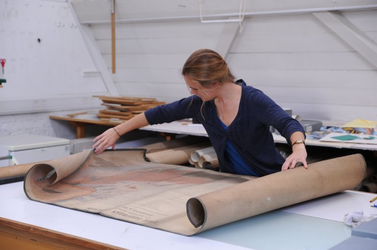
“At the start of my internship I worked in the Library archive with the more well-used material that is already collated and stored by author. But later I got the chance to sort through the archive of maps and charts that is hidden away in the old paper conservation studio.

Excitingly, the first thing we pulled out was a signed map by Roderick Impey Murchison, produced between 1831 and 1838, depicting the Silurian region and adjacent counties of England and Wales – the first geological map produced of that region!

Murchison was a Scottish geologist who in 1831 started studying the greywacke rocks underlying the Old Red Sandstone at the border of England and Wales. He subsequently grouped them into a new order of succession, which he called the Silurian System, a period lying between the older Ordovician and younger Devonian periods, and with its own distinct organic remains and unique rock formations.

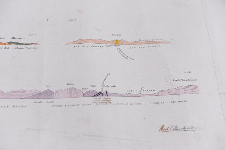
I later found three watercolours of numerous geological sections from the Lower and Upper Silurian, signed by Murchison but owned by William Buckland, Oxford’s first Reader in Geology.

dsc_64631
A watercolour signed by Murchison.

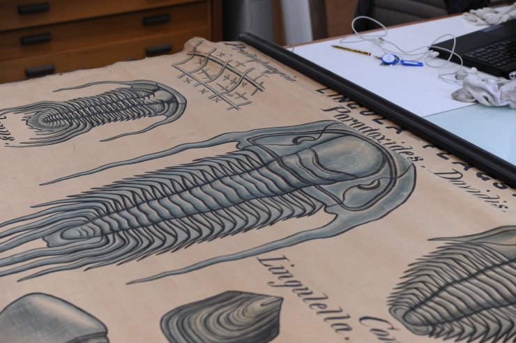
I’ve since encountered a whole array of maps of various parts of the world, from regional maps of the Nile basin in Egypt, to small parishes in the UK, through to maps of continents and the world, all dating between 1836 and 1924. Other finds in the archive have included numerous lecture diagrams depicting mainly fossil shells and trilobites from Silurian and Devonian rock deposits. These are in the style of English geologist John Phillips, who was William Buckland’s successor as Reader of Geology. Phillips may have used the displays as lecture aids during his time at the University, from 1853 to his death in 1874.

dsc_6473
A lecture poster depicting trilobites, in the style of, and possibly used by John Phillips, Reader in Geology at the University of Oxford, 1853-1874.

Most of the maps and charts are in need of some conservation care, including cleaning and storage in conservation-grade boxes. I have also been cataloguing them with descriptions so that they can be uploaded to the new content management system for later searching online. These descriptions include information on the author, date, and dimensions of an item, as well as an outline of its key features, language, location and condition.

However, the majority of the material has no associated authors or dates, so the next stage would be to trace their origins so that this information can be added.”

A job for another treasure hunter perhaps?

Cecilia Karlsson – EPA Cephalosporin Fund intern, 2014
Scott Billings – Public engagement officer