Writing from experience

The Museum’s building and collections provide inspiration for scientists and artists alike, often acting as a springboard for the creation of new work. Following a year here as one of three poets-in-residence, Kelley Swain returned to lead a session with Oxford Scholastica students, showing how museum objects can inspire creative writing.

by Kelley Swain

The experience of looking at the taxidermy Little Owl (Athene noctua) provided inspiration for Tallulah’s poem

Delving into the archives and behind-the-scenes stores, meeting researchers and conservators, and finding inspiration in the architecture, history, and collections were all part of my residency at the Museum during 2016. I’ve always written poetry inspired by the history of science and its fascinating objects, and I have come to appreciate museum objects not only as inspiration for my own poetry, but as teaching tools, or “object lessons” to inspire others.

It was lovely to be asked to lead a new series of these “object lessons” for a group of summer school students at Oxford Scholastica. Some of them had never encountered taxidermy, let alone a room full of articulated, stuffed, and preserved specimens. Awe abounded – both its wonder and, for some, its horror. It was a great opportunity to teach the students not only poetry, and why writing poetry inspired by museum objects can be moving, thoughtful, and important, but also to teach them about conservation and preservation.

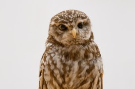
Here we share the work of 13 year old Tallulah Xenopoulos, who created this poem following an encounter with a taxidermy owl during the workshop:

Stupid dead owl.
The wooden door opens slowly, and, although there’s a green stone with bumpy edges and
shiny sides, a jar filled with silky insects and a board with beautifully painted butterflies.
Both your eyes land on the owl.
His feathers brush down his back and he stares down at his lightly spotted blanket where his
delicate legs connect and hatch onto the bumpy branch.
His eyes
And his beak
And legs
And nails
He stares at you almost like he knows what you’re thinking – which is dumb because he’s
dead – but he scares you and fascinates you at the same time.
A piece of dust has fallen beneath his eye and I bet he’d love to just brush it away, cause
he’s like that.
But also.
He’s an owl.
A stupid.
Dead owl.
With nothing but stuffed insides and scrawny legs.
And a heart. A dead heart which they slipped out and replaced with stuff.
-”do you think they stuffed him alive?”
The boy next to you whispers. You don’t reply. But the thought of death. And of his feathers
falling the second he felt the blood rushing through him go cold and dusty, travels across
your mind.
“Do you think he knew he was about to be?” you answer
Because the poor clueless animal looks as if he knew nothing.
knows nothing.

Kelley Swain’s own poetry from the Museum residency is featured in Guests of Time, a beautiful hardback volume edited by Prof John Holmes which features new work by John Barnie and Steven Matthews, alongside 19th-century poetry from writers linked with the early days of the Museum. Together, the poems in this anthology are a tribute to the Pre-Raphaelite origins of the Museum and a rejuvenation of its artistic legacy.

Mammoth tusks and cocktail sticks

By Pete Brown, Move Project Assistant

As part of the Museum of Natural History Move Project Team I have helped move and repackage tens of thousands of specimens since 2017, removing boxes filled at any time over the last 150 years from their old storage location in a deconsecrated church building near Oxford.

At our new facility we have been documenting and repacking the contents in new, clean containers and placing them in environmentally stable, safe warehouses specially adapted for museum storage.

Some objects are trickier to store than others. Things that are long, heavy, curvy and fragile are tricky. Mammoth tusks are long, heavy, curvy, and fragile. This means:

  1. They’re not going to fit in a normal box.
  2. They’re going to be difficult to move around.
  3. That beautiful curve will mean that all the weight of the tusk may be bearing down on just two small contact points where the tusk meets the storage surface.
  4. Because those points are fragile, they’re likely to get damaged.
A lot of weight can rest on small areas of the tusk, putting strain on the specimen and potentially causing damage

The tusk in this article is a prime example. The area nearest the camera in the photo above provided just a tiny point of contact with the floor and was very loose, almost to the point of detaching. It needed to be repaired, and stored in such a way that it wouldn’t get damaged again.

Pete Brown carries out delicate conservation work on the mammoth tusk

I filled some of the missing areas around the fragile area with an easily removable fine acrylic putty to prevent further movement and loss of the original material. A cotton tape sling helped to suspend the fragment in place during the work.

Thick plastazote provided a sturdy, slightly yielding bed for the tusk to lie on in storage, but to prevent the tusk from getting damaged again more needed be done to reduce the pressure on the points of contact.

The dark grey foam material, plastazote, is often used as a cushioned support for museum objects

I cut depressions into the plastazote where the tusk naturally lay to increase the total surface area supporting the weight of the tusk, and fixed plastazote wedges and supports in place with cocktail sticks to again increase the contact area and prevent movement. Cotton fabric ties, fed through slits in the plastazote, also helped to guard against unwanted movement.

Cocktail sticks: not just for cheese and pineapple

The repaired end of the tusk is now only supporting a fraction of the weight it used to, and once the tusk and the plastazote bed are placed into their new custom-made crate it will be ready for long-term, safe, damage-free storage!

The end of the tusk after treatment

To keep up with all the move project action, follow the museum hashtag #storiesfromthestore on Twitter @morethanadodo.


Bursting into life

By Ricardo Pérez-de la Fuente, Museum Research Fellow

One of the earliest and toughest trials that all organisms face is birth. In egg-laying animals, the egg shell that has protected the embryo during its early development ultimately becomes a hard barrier between the animal and its life out in the world. The bursting of the egg is literally a threshold moment, and there are many ways to crack an egg…

Some animals break the egg membranes using dissolving chemicals; others physically, mechanically tear their way through the shells. Among the latter, a great diversity of animals use specialised devices called egg bursters. These vary greatly among the many arthropods and vertebrates that use them, but perhaps the most famous example is the ‘egg tooth’ that is present on the beak of newborn chicks.

Four complete Tragychrysa ovoruptora newborns preserved together with egg shell remains and one egg burster. Modified from the open access Palaeontology paper.

My colleagues and I have found an exceptional fossil in 130 million-year-old Lebanese amber. Inside, trapped together are newborn larvae from Green Lacewings, the split egg shells from where they hatched, and the minute egg bursters that the hatchlings used to crack the egg. This is a first: no definitive evidence of these specialised egg-bursting structures had been reported from the fossil record of any egg-laying animals, until now.

The finding has been recently published as open access in the journal Palaeontology. Because multiple newborns were ensnared and entombed in the resin simultaneously, the fossil larvae have been described as the new species Tragichrysa ovoruptora, meaning ‘tragic green lacewing’ and ‘egg breaking’. A sad event, indeed, taking place in an ordinary day 130 million years ago in the Cretaceous forests of Lebanon, yet a happy circumstance now that we can take a privileged glimpse into the adaptations and behaviours of these fascinating tiny creatures.

The hatchlings from modern Green Lacewings open a slit on the egg with a ‘mask’ bearing a saw-like blade. Once used, this ‘mask’ is shed together with the embryonic cuticle and is left attached to the empty egg shell.

With the help of Amoret Spooner, Collections Manager at the Museum, egg clutches from modern green lacewings were found in the Museum collections. These eggs happened to have the intact egg bursters still attached to them, and proved to be crucial to understand that we had the same structures preserved in the amber together with the newborn larvae.

Two Tragychrysa ovoruptora newborns preserved together with egg shell remains and two visible egg bursters (right inset). Modified from the open access Palaeontology paper.

Green Lacewing larvae are small predators that often carry debris as camouflage, using their sickle-shaped jaws to pierce and suck the fluids of their prey. Whereas the larvae trapped in amber differ significantly from modern-day relatives, in that they possess long tubes instead of clubs or bumps for holding debris, the studied egg shells and egg bursters are remarkably similar to those of today’s green lacewings.

The larvae were almost certainly trapped by resin while clutching the eggs from which they had freshly emerged. Such behaviour is common among modern relatives while their body hardens and their predatory jaws become functional. Indeed, the two mouthparts forming the jaws are not assembled in most of the fossil larvae, which indicates, together with the large relative size of the head and legs, that they were recently born.

Detail of a head with the jaws still dislodged, indicating that the larva was recently hatched when it was ensnared by amber and the jaws had not yet had time to fully assemble.

It may seem reasonable to assume that traits controlling a life event as decisive as hatching would have remained largely unchanged during evolution. In fact, we see in very closely related insect groups different means of hatching that can entail the loss of the egg bursters. So the persistence of a hatching mechanism in a given animal lineage through deep time can’t be determined without direct proof from the fossil record.

Reconstruction of two Tragichrysa ovoruptora newborns clutching the eggs from where they recently hatched, moments before they were trapped by resin. Larvae colour and egg stalks are conjectural. Extracted from the open access Palaeontology paper.

This new discovery shows that the mechanism green lacewings use to crack the egg was already established 130 million years ago. Overall, it represents the first direct evidence of how insects hatched in deep time, egg-bursting their way through into life.

*

The hatching mechanism of 130-million-year-old insects: an association of neonates, egg shells and egg bursters in Lebanese amber by Ricardo Pérez-de la Fuente, Michael S. Engel, Dany Azar and Enrique Peñalver is published as open access in Palaeontology this month.

Imagining lost worlds

Earlier this year University of Plymouth illustration student Rachel Simpson teamed up with our research fellow Jack Matthews to ‘bring the oldest multi-cellular organisms back to life’. Rachel tells us about the process of working with some of the most ancient fossil material and reveals the results of her illustrations and modelling.

Illustration by Rachel Simpson, created in collaboration with the Museum

In August 2018 I was lucky enough to travel to Newfoundland, Canada with Dr Jack Matthews to learn about and illustrate some of the extraordinary fossils found there. A highlight of the trip was going down onto the fossil surface – known as the MUN surface – to look at examples of organisms such as Beothukis, Charnia and Primocandelabrum, all of which date from the Ediacaran period, over 550 million years ago.

The MUN surface is the location of the fossils that I had worked on for my university project. I had spent the previous months sketching, drawing and bringing these organisms back to life from silicon casts, so it was amazing to be able to see the real specimens in situ and to sketch from the fossil surface.

Sketching directly from the fossils also provided a new challenge as I was unable to control factors such as the lighting, which is crucial to seeing the fossils clearly. Nonetheless, I learnt a lot about drawing on location.

Sketching at the fossil surface

While visiting Port Union I was able to use some of the old printing presses held by the Sir William F. Coaker Heritage Foundation to create work inspired by the fossils I had seen in the surrounding area. I love using printmaking in my own illustrative practice so it was a great experience to get to use these old presses (image at top of article).

We also had the chance to give a radio interview and talk to the Port Union community about the work that Jack and I had done, showing how science and art can work together.

On my last day in Port Union I was invited by a local potter to make some ceramic representations of the fossils I had been drawing there. I created models of Fractofusus and Aspidella, and discovered that re-imagining something in three dimensions is a very different process to recreating it as a drawing.

Rachel created ceramic representations of some of the Ediacaran organisms

For the final three days of the trip we relocated from Port Union to Trepassey to visit the Mistaken Point UNESCO World Heritage Site. Here, I saw the highly preserved Fractofusus specimens and made some more sketches. Using a small hand lens I was able to draw all the details that are invisible to the naked eye.

Using a hand lens allowed Rachel to pick out details in the Fractofusus fossil

Drawing on location in Canada provided a better idea of the organisms in relation to other surrounding organisms, something that is more obscure when working from museum specimens. This definitely informed my practice and meant that artwork created after the trip was more representative of the science.

When I returned to England, I created some new prints inspired by my time in Newfoundland, the fossils that I saw, and the printing process I was able to use in Port Union.

A set of prints made by Rachel based on her work in Newfoundland

Crafty camouflage

Last week we brought you snails that attach all manner of pebbles, fossils, corals and shark teeth to their shells. Today we give you a newly-discovered fossil green lacewing larva that attached pieces of soil to its body as an act of camouflage. Our research fellow Ricardo Pérez-de la Fuente, lead author of the new paper, explains…

Visual camouflage is one of the most successful survival strategies in nature. Camouflaging is usually defensive, allowing animals to be left unnoticed by their predators, but it can also be used aggressively by predators themselves to approach their prey undetected.

Some camouflaging animals can actively change their colouring to match that of the background ‒ a technique called crypsis. Others can make their bodies resemble elements of the environment, such as leaves or twigs, which is called mimicry.

Italochrysa italica, an extant green lacewing larva carrying a dense debris packet made of soil fragments. Taken from the open access publication Tauber & Winterton, 2014.

Yet another approach to camouflage involves collecting diverse materials from the environment and incorporating them on the animals’ bodies in order to better blend with the surroundings. This is known as debris-carrying, trash-carrying, or decoration, and it can be found across a wide variety of animals including sea urchins, gastropods, and arthropods, such as decorating crabs, or sand- and mud-covering spiders.

My colleagues and I have just published the discovery of a fossil green lacewing larva, pictured at the top of the article, that has been preserved carrying bits of soil that it used for camouflage and physical protection. It’s a new larval species just 1.5 mm in length, and is preserved in Early Cretaceous Lebanese amber. We have named it Tyruschrysa melqart after the Phoenician city of Tyre and its tutelary god Milk-Qart (if you want to learn the reasons behind this name check out our open access paper!).

Interpretative drawing of Tyruschrysa melqart: body in grey, ‘tubes’ with setae coloured according to which body part they are attached to, and soil debris in brown.

Green lacewing larvae are active predators that eat other insects such as aphids, using sickle-shaped ‘jaws’ to pierce their prey, suck out their fluids and liquefy their tissues; eating is easier when there is no need to chew! Some green lacewing larvae are debris carriers, entangling all kinds of debris among their velcro-like ‘hairs’ called setae, which extend from relatively short ‘bumps’ on their backs. This debris is carefully selected and gathered with meticulous head and body movements to form a so-called debris packet on the back of the insect.

‘Tubes’ bearing setae of Tyruschrysa melqart, with detail of their mushroom-shaped endings (bottom), used for anchoring bits of soil.

The new fossil and similar ones described from younger Cretaceous ambers differ from modern relatives because instead of short ‘bumps’ with setae on their backs they have relatively long ‘tubes’, giving them a bizarre appearance.

These tubes have setae with mushroom-shaped endings of a kind never seen before in extinct or living green lacewing larva species. The mushroom-shaped ending is a special adaptation to anchor debris, which in the case of Tyruschrysa melqart are fragments of soil.

Hallucinochrysa diogenesi, another Cretaceous green lacewing larva bearing long ‘tubes’ with setae on its back, but carrying a debris packet made of plant hairs (trichomes). Preserved in Spanish amber (105 million years old).

It was already known that Cretaceous green lacewing larvae like Tyruschrysa had long tubes on their backs and that they collected plant hairs and other plant material to construct their packet of debris. But thanks to the new discovery we now know that these immature insects also used bits of soil, and that in the deep past debris packets were probably as diverse as those we see today.

Green lacewing larvae have been gathering debris to camouflage and protect themselves for about 130 million years, giving rise to the different body adaptations we see amongst these fascinating tiny collectors.

‘A soil-carrying lacewing larva in Early Cretaceous Lebanese amber’ Ricardo Pérez-de la Fuente, Enrique Peñalver, Dany Azar and Michael S. Engel is published as open access in Scientific Reports this month.

Restoring the Great Lizard of Stonesfield

by Paul Smith, Museum director

One of the unusual things about the collections in the Museum is that some of the specimens date back hundreds of years, and so have been researched by generation after generation. Sometimes these specimens have been damaged and repaired, and in some cases this has happened many times, leaving a complex history of research and conservation.

One high profile example is the type specimen of the theropod dinosaur Megalosaurus bucklandii – the world’s first scientifically described dinosaur. This specimen itself is the lower jaw, pictured above, which has been in the collections of Oxford University since 1797 at the latest.

Working with the Centre for Imaging, Metrology, and Additive Technology (CiMAT) at WMG, University of Warwick, we have been unraveling the conservation and repair history of the fossilised jaw using an innovative combination of modern technologies.

Identification of repair using X-ray computed tomography (XCT ) from the Megalosaurus bucklandii type specimen. The two colours indicate two different types of plaster material. Scale bar is 10 cm.

Earlier studies had mapped the presence of plaster used for repair, but X-ray CT scanning of the type used in medical procedures rapidly revealed a number of different phases of repair. In each of these repairs the plaster was of different composition and was used in different places.

One type, shown in red on the image above, was used to infill fractures and to make the specimen more robust; a second type, shown in green, was used to repair the teeth and, in some cases, to recreate the teeth. Interestingly, the extent of plaster revealed by the CT scanning is actually less than previously interpreted with the naked eye.

The two types of plaster were then analysed chemically to better understand their historical use, revealing quite different compositions. The more abundant ‘red’ plaster is mixed with quartz sand and calcite grains, possibly from the rock matrix surrounding the fossil, to make it look more similar.

Carbon is also abundant and grains rich in lead are present. Carbon is not common in the rock itself, and the carbon in the plaster has probably come from a varnish such as shellac being used to coat the repair.  The presence of lead was more puzzling. Further analysis eventually showed that the grains were made of red lead – lead tetroxide – which was used historically as a pigment in paint. The red lead in the repair may have been used to colour the plaster, but it may also have been applied to make the density of the plaster more similar to that of the fossilized bone, and so replicate the weight of the specimen better.

Reconstruction of Megalosaurus bucklandii by Julius T. Csotonyi

The second type of plaster, used to repair the teeth, lacks the lead of the first type but contains barium. Barium hydroxide was often used as a consolidant and sealant for plaster, which would explain its use here.

Having a full understanding of the repair history and the position and extent of plaster helps us in a number of ways. It allows researchers to understand which parts of the lower jaw are original and anatomically reliable, and it helps the curators and conservators to know which parts of the specimen are more fragile during handling and display.

By combining cutting-edge scanning technologies with heritage material we are able to shed new light on the conservation history, and future, of important specimens such as Megalosaurus bucklandii.

Read the full paper here.