Crab in the lab

After_small

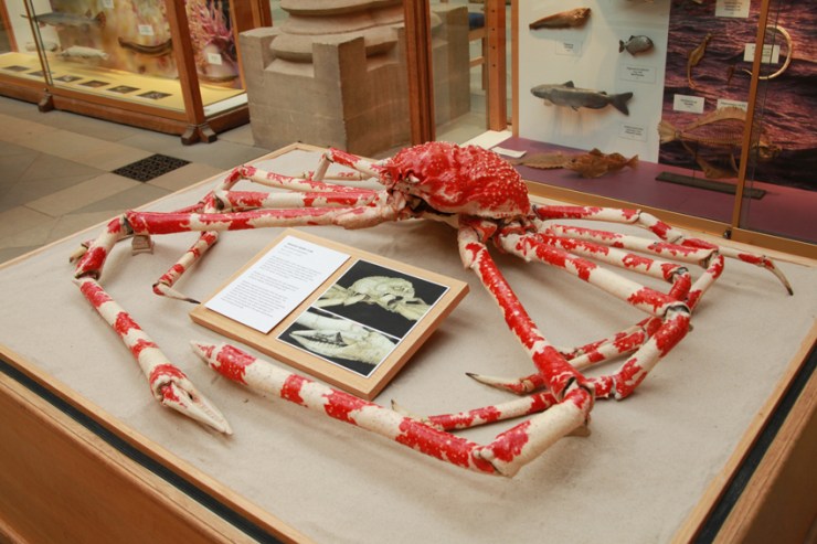
One of the most loved specimens in the Museum is the enormous Japanese Spider Crab. It’s been on display for over 100 years, so it’s unsurprisingly shown serious signs of deterioration. In July, staff in our Life collections decided that the crab should come off display and undergo conservation treatment. Bethany Palumbo, Conservator in Life Collections, took charge of this famous specimen.

The spider crab looking washed out after 100 years on display

The most obvious damage was the loss of colour – the natural carotenoid pigments had completely faded due to decades of continuous light exposure under the glass roof. However, once it was taken into the laboratory for a closer look, Bethany soon realised that there were actually many areas that were fake, composed of old materials such as acidic cardboard, newspaper and even carved wood.
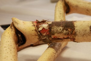
Some of the old filler material in a leg

These restoration efforts were causing more harm than good, deteriorating and damaging the natural shell material. The whole specimen was loosely held together with animal glue, PVA adhesive and, in some areas, tough wire which was cutting through the shell.

The first step for our conservator was to check the Museum database for information about the specimen, such as when it was donated and by whom. But unfortunately the specimen has no record, nor is it accessioned into the Museum’s collection. Although frustrating, this was important information. As it had no scientific data, Bethany could give this specimen more extensive conservation treatment, without compromising its scientific or historic integrity.

Bethany decided that treatment would consist of cleaning, the removal and replacement of old, deteriorating fill material and the restoration of colour to the shell, making the specimen true to life. These treatments, with the exception of the cleaning element, would be completely reversible.
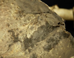
Dirt build-up on the underside of the shell

Work began by taking the specimen apart to clean and treat each section. Sections were gently vacuumed and a moist cloth used to wipe away 100 years of embedded grime. Bethany removed old fill material, softening it with water vapour to allow it to be easily peeled away.

Newly filled and coloured leg

The next task was to create replacements for the missing sections. Bethany used a combination of acid-free tissue and closed-cell polythene foam. Intricate areas like the claws proved more challenging. Replacements were sculpted free-hand from Plasticine, moulded in silicone and finally cast in Jesomite composite plaster. They now look pretty close to the real thing and are a big improvement on the old wood and paper versions.

Replacement parts made from Jesomite plaster

Before it was ready to go out on display Bethany replaced the faded colour. Japanese Spider Crabs are bright red and white in life, but ours had become washed out beige.
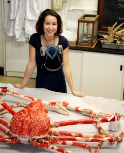
Conservator Bethany with the finished Spider Crab

Photographs of Spider Crabs were used as a reference for the colours, and Bethany also spoke to crustacean experts in the Museum to make sure it was accurate. She used an airbrush and acrylic inks, selected for their high UV resistance. The shell was coated with a barrier layer to allow the ink to be removed in the future, if needed.

Airbrushing the specimen was the most time consuming element, as it required multiple layers and various brushing techniques to make the crab look true to life. Once completed, Bethany gave the crab a final protective coating, providing good water resistance, ready for the next time it needs a good clean!

Rachel Parle, Interpretation and Education Officer
On the wing

Jones 2

Since the 1920s the Museum has had in its care an original, unpublished manuscript containing over 1,500 beautifully detailed and colourful paintings of butterflies and moths. Known as Jones’ Icones, this one-of-a-kind work was created in the late 18th century by amateur Lepidopterist and retired London wine merchant, William Jones.

The paintings depict over 760 butterflies and moths from the collections of some of the most eminent naturalists in London at that time, including the founder of the Linnean Society, Sir James E. Smith and entomologist Dru Drury, as well as Jones’s own specimens.

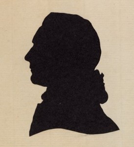
A silhouette of William Jones

In addition to being a beautiful work of art, Jones’ Icones is also an extraordinarily important document in the history of entomology and insect collecting in Britain. At the time Jones was making these paintings, many of the specimens he was depicting were being brought to Europe and described for the first time, most notably from Australia and the Oceanic region. Jones meticulously recorded these specimens through his paintings, and his work remains the only record of many of these important collections, a large number long-since destroyed, lost, or divided among private collectors.

The Icones was also consulted by a student of Linnaeus, Johann Christian Fabricius – the man credited as the first to describe over 10,000 insects. It is believed that Fabricius named over 200 species from the images in the Icones, citing Jones’ work in his publication Entomologica Systematica in 1791.

Large Copper from Jones Collection at the Museum
Large Copper butterflies (Lycaena dispar) from Jones Collection at the Museum

In spite of this manuscript’s huge importance to the history of entomology in Britain, it has never been made available beyond the reading room of the Museum’s archive. So now, after almost a century of failed or abandoned attempts, Jones’ Icones is available for all to see!

A plate from the Jones' Icones
A plate from the Jones’ Icones

As part of an HLF funded project, Flying Icons, which has been running since April 2013, all six volumes of Jones’s Icones have been digitised and made available online. A website at www.jonesicones.com has also been developed in order to promote this amazing manuscript and the related collection here at the Museum.

This website also serves another very important function: to solicit help from keen amateurs and experts worldwide to help identify all the species represented in Jones’ Icones. Identification is the first step in tracking down extant specimens of some of Jones’ paintings. It may even change our understanding of the history of the science of entomology. Can you help? If you think so, request an account and we’ll set you to task!

If you’d like to read more about Jones’ Icones – and see some large scale prints – then head to the current exhibition in the cafe area of the Museum.

Kate Santry – Head of Archives and Library

Jones

Every breath you take

IMG_3738

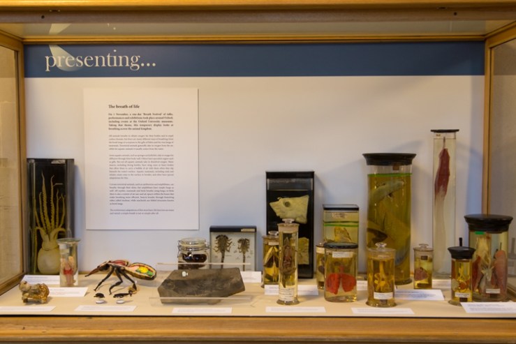
Tomorrow afternoon the Museum will host talks, tours and a dance performance as part of the Breath Festival, a unique series of events coordinated by the Oxford University Hospitals Artlink programme. To coincide with the festival we have put together a special display in our changing Presenting… case, all about breath and breathing across the animal kingdom.

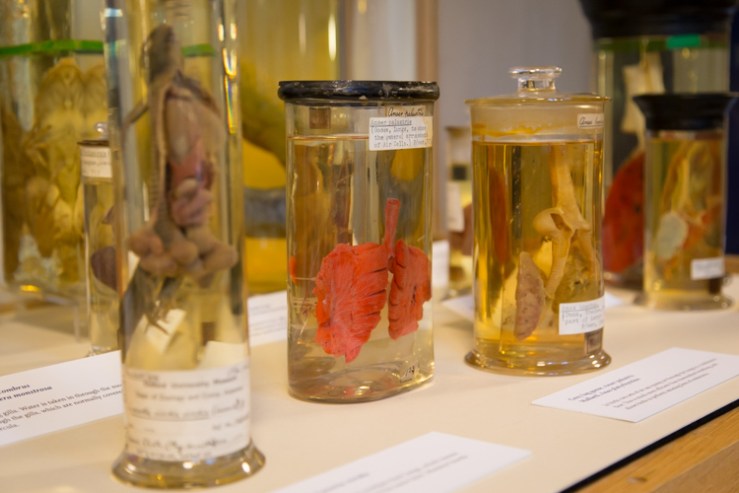
There’s something of the Halloween macabre about the display too, with its pink-coloured lungs and eviscerated bodies suspended in spirit. Here’s a taster of the display, but to see the full selection head down to the Museum either for the Breath Festival tomorrow, Saturday 1 November, or at any time during the rest of the month.

Lungs of a lizard, goose and duck.
Lungs of a lizard, goose and duck

The breath of life
All animals breathe to obtain oxygen for their bodies and to expel carbon dioxide, but there are many different ways of breathing: from the book lungs of scorpions to the gills of fishes and the true lungs of mammals. Terrestrial animals generally take in oxygen from the air, while for aquatic animals it usually comes from the water.

Crocodile and alligator lungs

Some aquatic animals, such as sponges and jellyfish, take in oxygen by diffusion through their body wall. Others have specialist organs such as gills. But not all aquatic animals take in dissolved oxygen. Many insects, including diving beetles, have wing cases or hairy bodies that allow them to carry a bubble of air with them when they dip beneath the water’s surface. Aquatic mammals, including seals and whales, must come to the surface to breathe, and often have special adaptations for this.

Certain terrestrial animals, such as earthworms and amphibians, can breathe through their skins, but amphibians have simple lungs as well. All reptiles, mammals and birds breathe using lungs; in birds there is also a system of air sacs and air spaces within the bones that make breathing more efficient. Insects breathe through branching tubes called tracheae, while arachnids use folded structures known as book lungs.

The evolutionary adaptations of this most basic life function are many and varied: a simple breath is not so simple after all.

Visitors pick out their favourite specimen

Scott Billings – Public engagement officer

Plight of the bumblebee

nhm16

The ecological importance of bumblebees has become more widely appreciated in recent years, thanks to environmental campaigners and reports of species decline, and even some extinctions, in the UK.

To look at this issue, we have recently teamed up again with arts-science organisation Pale Blue Dot, which is launching a new research project to investigate why some species of bumblebee are declining and to raise awareness about the ultimate impact this has on people.

Here, Pale Blue Dot co-founder Jane King explains how the Bees & Weeds project brings together art students, public engagement, the Museum’s collections and a leading bumblebee scientist…

*

On 9 September we launched our latest project – Bees & Weeds – with the Museum of Natural History, building on our previous collaboration for the Lost & Found exhibition. We were joined by over 50 art students from Banbury & Bicester College to highlight the plight of the bumblebee, revealing how its decline is impacting everything from what we eat to where we live and work.

Amoret Spooner displays drawers from the collections

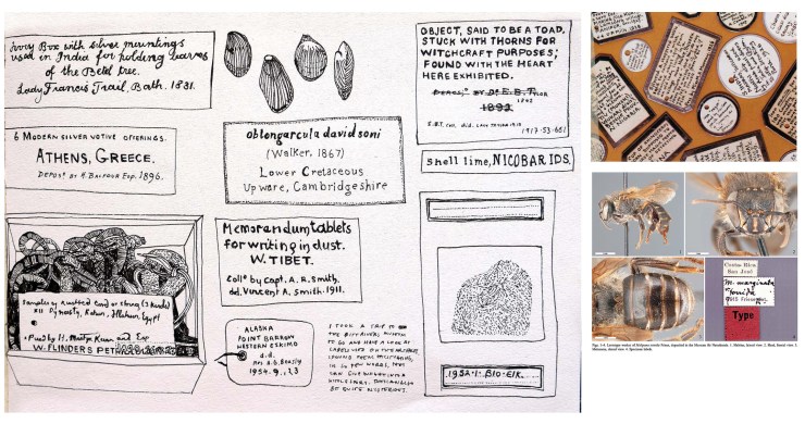
The students spent some time looking at methods of insect labelling and notation, before heading behind the scenes with entomologist Amoret Spooner to the Huxley Room, the location of the Great Debate on Darwin’s Theory of Evolution, which took place in 1860.

Amoret provided an insight into taxonomy – the science of species classification – as well as her work on the conservation of specimens. We visited the huge archive of bee specimens and learnt about some of the research that scientists are currently carrying out on UK bumblebee species to help prevent further decline.

Student sketches of labelling and notation

During spring 2015, the art students from the Banbury & Bicester College, as well as students from Oxford Brookes University, will make and install hundreds of cycle seat covers on bikes in and around the Oxford city. The seat covers will carry messages about bumblebee decline in the UK countryside, showing how much we depend on their pollination services, which far outstrip those of the honeybee in their value to UK food production.

We are also working with Professor Dave Goulson from the University of Sussex, one of the world’s most important bumblebee scientists. He will present his research showing how well bumblebees are doing in gardens compared to the countryside, as well as the optimum range of flowering plants needed to help them thrive. Dave’s book, A Sting in the Tale, is already a best-seller, and the sequel – A Buzz in the Meadow – was published on 4 September.

Dave will also be speaking about his new book at the Museum on Thursday 9 October at 7pm. Book your tickets for that via Waterstones here.

Artwork from the Bees & Weeds project, together with cycle seat covers and bike paraphernalia, will be on show and for sale in the Old Fire Station in Oxford from next spring. If you cycle in Oxford, you may be lucky enough to receive one!

Pale Blue Dot is an arts-science organisation helping scientists to communicate their research to the public. It promotes an interdisciplinary approach to learning, living and working through exhibitions, publications and happenings.

Jane King – co-founder, Pale Blue Dot

Summer Swifts

Judith Wakelam 04
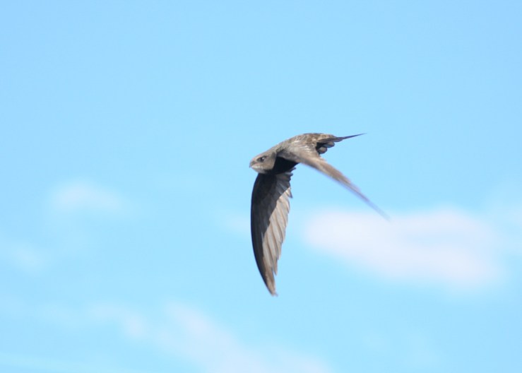
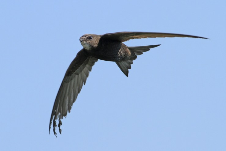
Mastery of the Skies by Judith Wakelam

Hopefully many of you have been able to visit the Museum over the summer to admire the incredible images in the Wildlife Photographer of the Year exhibition. If you haven’t yet seen the inquisitive lion or the mysterious elephants, don’t leave it too long; it’s only on display until 22nd September.

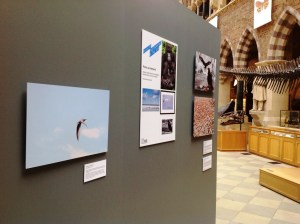
Mastery of the Skies on display in the Museum

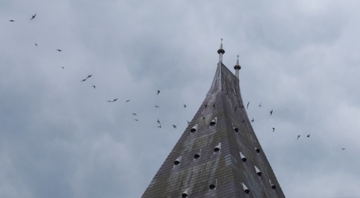
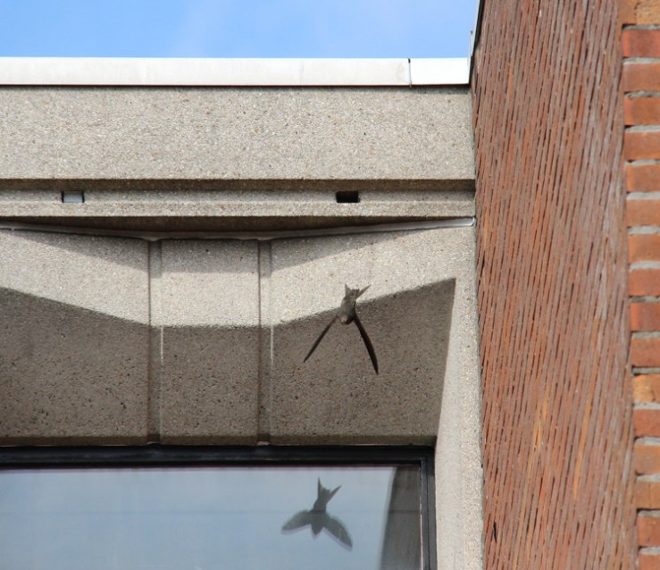
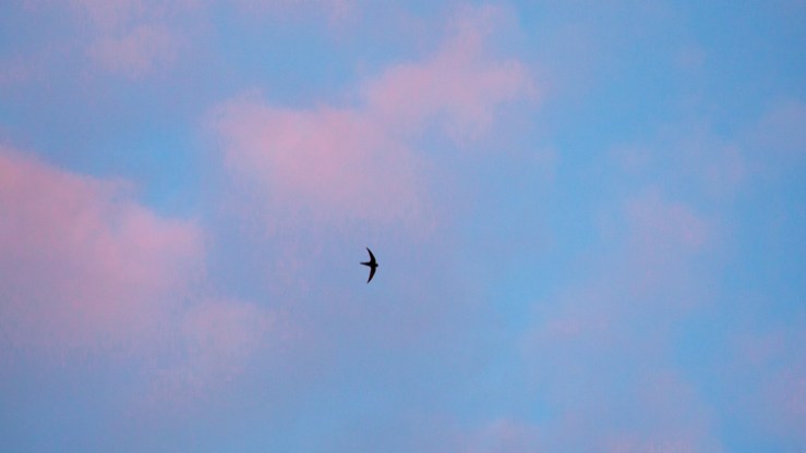
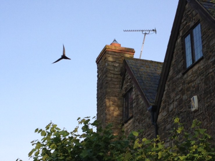
Today we’ve had a new addition to the spectacular collection of photographs. Mastery of the Skies by Judith Wakelam (above) will sit alongside these international competition winners. Judith’s photograph was chosen as the winner of the Museum’s own Summer Swifts competition, a challenge inspired by the display of the WPY exhibition here.

Photographing swifts is particularly difficult – they are small birds which fly very quickly and almost never stop to rest. Although the competition received some excellent entries, the judges felt that Judith Wakelam’s image really captured the character and dynamism of a swift in flight, showing some motion but still retaining enough clarity to easily see the bird’s face.

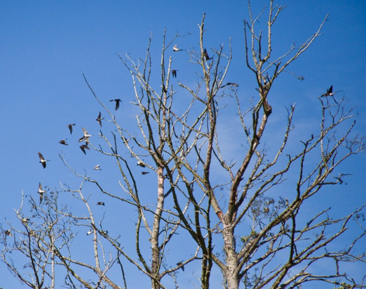
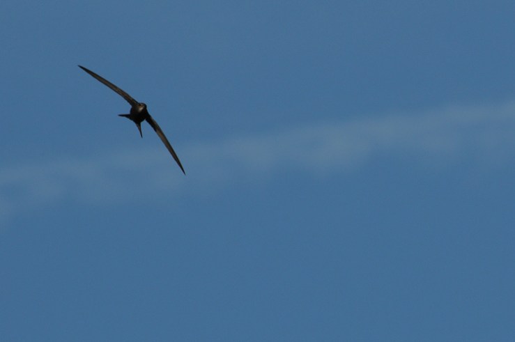
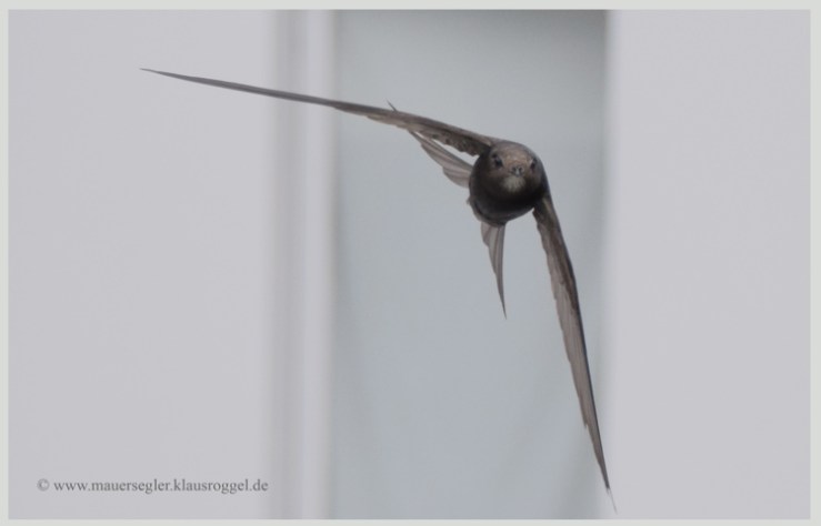
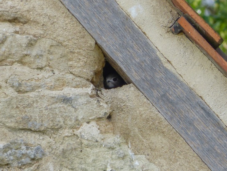
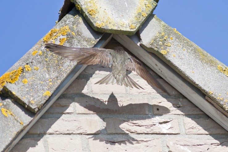
Here are the other Summer Swifts competition entries, so you can see for yourself how high the standard was.

Mark Garrett
Mark Garrett
Martine Tenret 2
Martine Tenret
Tom Nicholson-Lailey
Neil Downing 3
Neil Downing
Nick Owen
Nick Owen
Stephen Powles
Stephen Powles
Klaus Roggel
Klaus Roggel
Chris Powles
Chris Powles
Maciej Szymański 1
Maciej Szymański
Gordon Bowdery
Gordon Bowdery
Henk Haans
Henk Haans
Ingolf Grabow
Ingolf Grabow

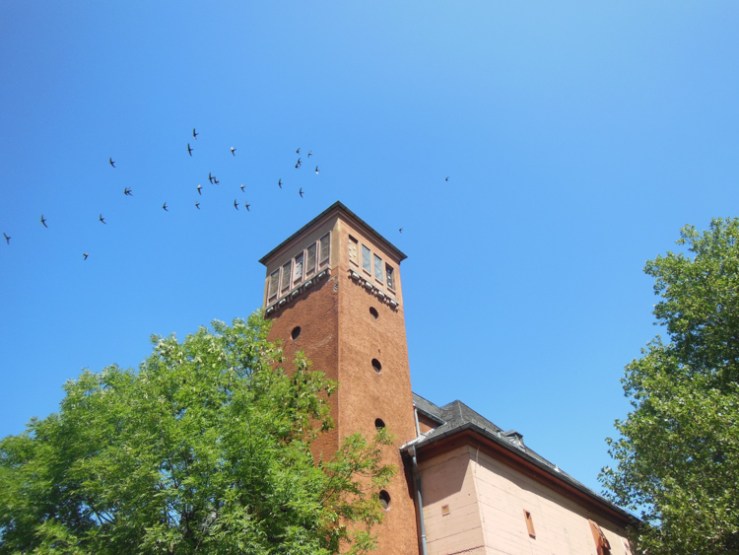
The Museum’s own colony of swifts has had a good summer and there’s now just one little chick left to fledge. We’ll all miss their distinctive screech as they soar around the Museum tower. Fortunately, Mastery of the Skies will keep the memory of high summer alive in the museum well into September.

Rachel Parle, Interpretation and Education Officer

Presenting… Dr Hilary Ketchum

IMG_2219

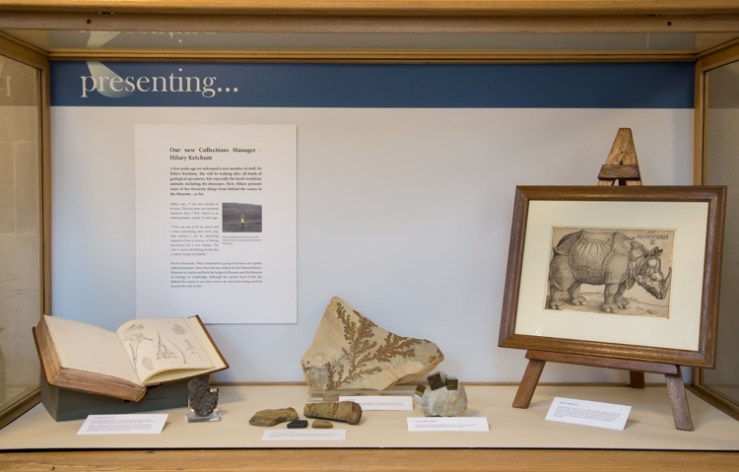
A few weeks ago we welcomed Dr Hilary Ketchum as our new collections manager for the geological collections. Hilary will be looking after all kinds of specimens, but especially the fossil vertebrate animals, including the dinosaurs. To welcome her, and to announce her arrival to the public, we have handed over our regularly-changing Presenting… display so that Hilary can exhibit some of her favourite things (so far) from behind the scenes in the Museum.

Hilary looking for plesiosaurs in the Oxford Clay on a rainy day. She’s never found one.

For her doctorate, Hilary researched a group of Jurassic sea-reptiles called plesiosaurs. Since then she has worked for the Natural History Museum in London and both the Sedgwick Museum and the Museum of Zoology in Cambridge. Although she spends most of the day behind the scenes in our store rooms she also loves being involved in activities and events.

I am very excited to be here. This has been my favourite museum since I first visited as an undergraduate, nearly 15 years ago. I love my job as it’s so varied and I learn something new every day. One minute I can be answering enquiries from scientists, or finding specimens for a new display. The next I can be identifying fossils that a visitor found on holiday.

A few of Hilary’s selection of specimens are include here. To see the full display, look for the Presenting… case just to the right of the Welcome Desk near the entrance to the Museum. An online archive of the Presenting… series is also available on our website.

IMG_2222
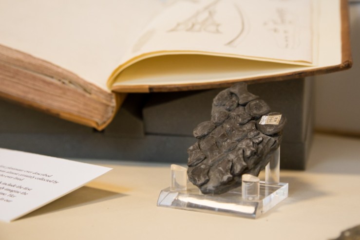
The first plesiosaur – Part of a flipper from the first plesiosaur ever described scientifically. It was almost certainly collected by Mary Anning, one of the greatest fossil-hunters who ever lived.
IMG_2223
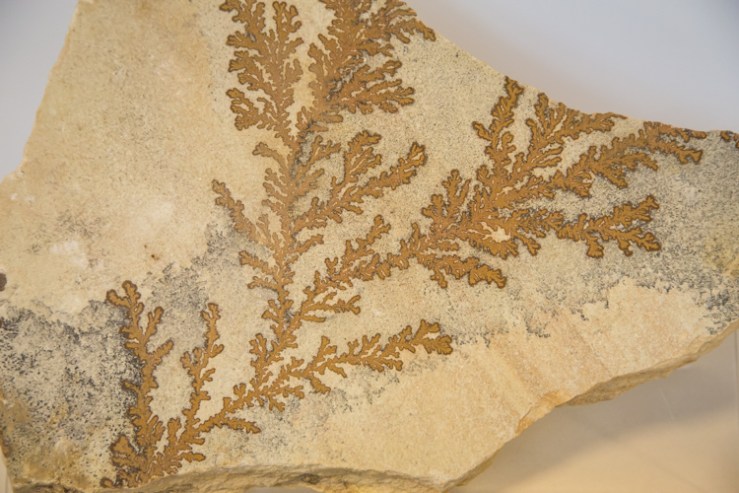
Dendrites – This may look like a fossil plant but it is actually a form of mineral growth called a dendrite. This type of crystal growth can also be found in snowflakes.
IMG_2220
Cubic pyrite crystals – This specimen of “Fool’s Gold” is from Spain. “I find it amazing that something so straight and orderly can arise in nature,” says Hilary.

Scott Billings – Public engagement officer