The winning brainwave

If you could create an experiment to learn more about the human brain, what would you investigate? We posed this question in our Big Brain Competition last year, as part of the Brain Diaries exhibition with Oxford Neuroscience, and received a whopping 800 entries!

For the competition, Oxford University neuroscientists offered people the chance to use the state-of-the-art MRI scanner at Wellcome Centre For Integrative Neuroimaging at the John Radcliffe Hospital to investigate a burning question about the brain. We had ideas from the young and old, and by visitors from all around the world suggesting brilliant questions and some fascinating experiments.

Memory_fMRI
Functional MRI image of the human brain using the MRI Scanner

To judge all the ideas, entries were split into categories: feasible experiments, unfeasible experiments, under 18s, and questions about the brain. WIN researchers compiled a long-list for each, which was ranked by a panel of neuroscientists and people from the museum to reach the eventual winners.

Sadly, only one experiment could be carried out, so an overall winner was picked from the ‘feasible experiments’ category. The winning experiment was suggested by Richard Harrow, who wanted to understand how the brain identifies voices.

A person is put in the MRI scanner with headphones on.  They are shown a photo of a person familiar to them, either a friend, family member or celebrity.  Then, in their headphones they are played the voice of a person, but the voice is either sped up or slowed down.
They are required to say whether the face on the photo matches the voice they have heard. What happens in the brain when this confusion of audio and visual information is occurring? Will the brain find a way to identify the vocal signature of the voice, even if distorted, and be able to say with conviction if the photo and the voice are a match?
– Richard Harrow, winning competition idea

IMG_4498 (2)
Competition winner Richard Harrow was interviewed alongside neuroscientist Dr Stuart Clare during the live streaming of the experiment

On the day of the experiment, the winners and runners-up headed over to the WIN Centre to watch Richard’s winning experiment being conducted. The experiment was streamed live by Oxford Sparks and we had a clear result from the test, as neuroscientist Dr Holly Bridge explains:

The scans show that when you’re getting information that corresponds in both your auditory and your visual system you get a boost in your brain activity. We can detect that the brain does respond differently depending on whether or not you can match the face with the voice; it clearly has a lot to do with expectation.

Stuart and Holly
The brains behind the Big Brain Competition, Dr Holly Bridge and Dr Stuart Clare explained the results of the experiment on a Facebook Live stream

The scientists also wanted to answer as many of the other great brain questions as possible. So a series of articles picks out some of the broad themes in the competition ideas, including lifestyle, muscle memory and stress. Researchers also answered more big questions live on Facebook during this year’s Brain Awareness Week.

We sorted the many entries in the Big Brain Competition into themes such as vision, lifestyle, and language

Thank you to everyone who suggested an experiment or asked a question; it made for a fascinating conclusion to the Brain Diaries exhibition, and has definitely increased the amount of brain activity from staff across the Museum and Oxford Neuroscience… if only there was an MRI scanner for us to see it!

IMG_3989
Winners of the Big Brain Competition at the Museum of Natural History with the neuroscientists. Left to right: James, Holly, Stuart, Misha, Richard, Lily, and Heidi

You can still get involved with the Big Brain Competition by trying the winning experiment at home.

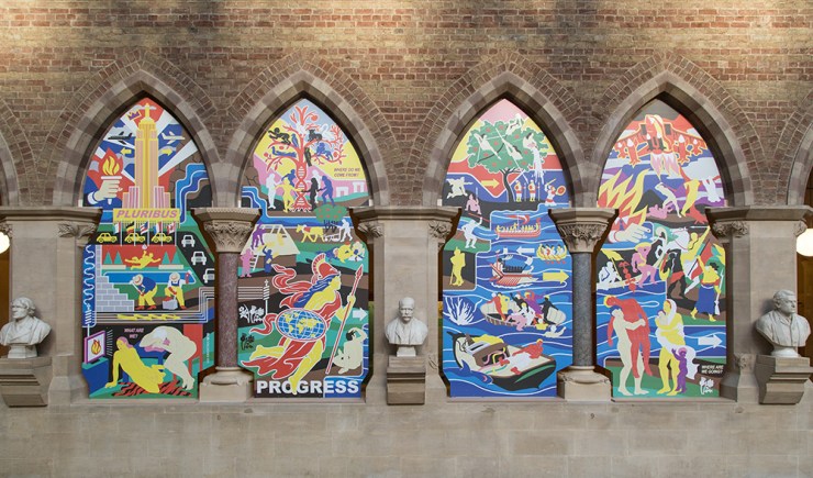
 

Where Do We Come From? What Are We? Where Are We Going?


These are some of the big questions asked in our current special exhibition, Settlers. It’s also the title of a new artwork by Ian Kirkpatrick that has just been commissioned by the Museum.

You may remember, back in July we put out a call for artists to respond to the main themes of the upcoming Settlers exhibition. We received an incredible response, with almost 100 proposals, so needless to say we were spoilt for choice! After several rounds of shortlisting, discussion and deliberation, we chose Ian Kirkpatrick, a Canadian artist now based in York.

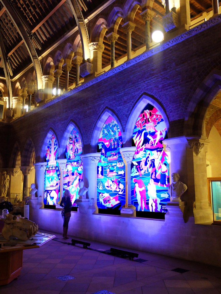
Lit up for the Settlers exhibition launch.
Credit: Ian Kirkpatrick

We were excited by Ian’s bold iconography and references to the history of art and design, while using shapes and colours usually seen on contemporary street signage. His approach to the themes and issues around migration, genetics and settlement were innovative and brave. We also couldn’t wait to see how his work would look in our Victorian neo-Gothic building.

Ian working in his studio. Credit: Ian Kirkpatrick

Over a period of four months, Ian researched, planned and created his spectacular final piece Where Do We Come From? What Are We? Where Are We Going? Here he explains a little about his artistic process:


Most of my projects begin with a period of research – often looking at historical events or interesting facts related to the brief. I often sketch out a very rough layout of the design in my notebook, then create the actual artwork directly onto the iPad or computer. Because I use vector-based software, I can easily rearrange or modify graphics – so the design is constantly shifting until the artwork is finished.

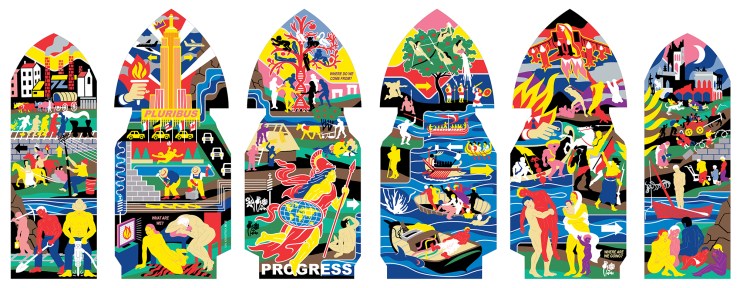
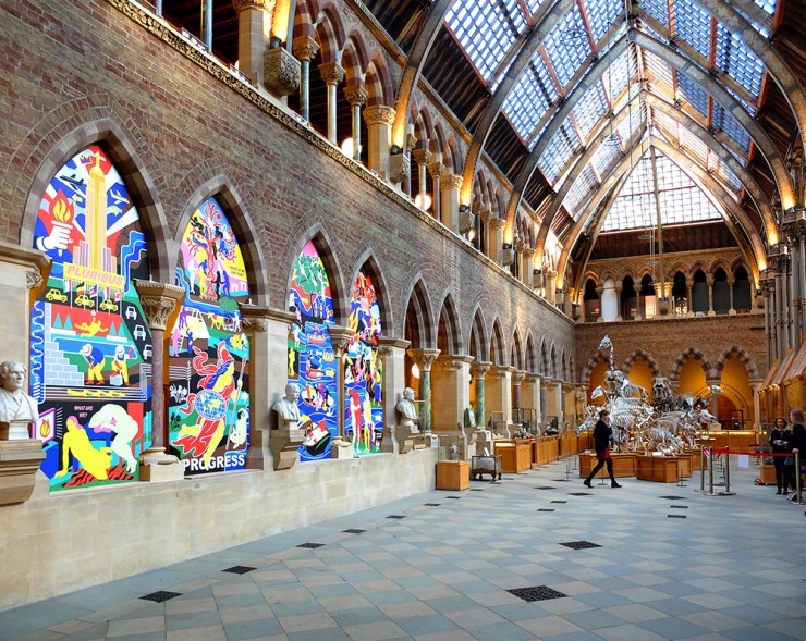
Ian Kirkpatrick’s final design. The two smaller panels (L and R) can be seen on display in the Settlers exhibition gallery.

Ian created a series of six panels that explores the social and natural causes behind human migration, both in ancient times and in the present day. It presents historical and modern peoples moving across a landscape in response to conflict, climate change and urbanisation, and remixes imagery from classical paintings alongside iconography from Great War postcards, Roman coins and the Bayeux Tapestry.

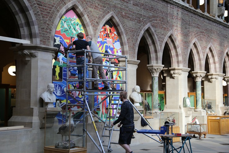
Ian, Peter Johnson and Adam Fisk installing the main panels of the artwork.

Of course, in a building like ours, the installation of such a large, bold piece of work would never be easy. Peter Johnson, the Museum’s Building Manager, came up with an ingenious solution to hold the panels into the arches, without damaging the masonry by drilling or glueing.

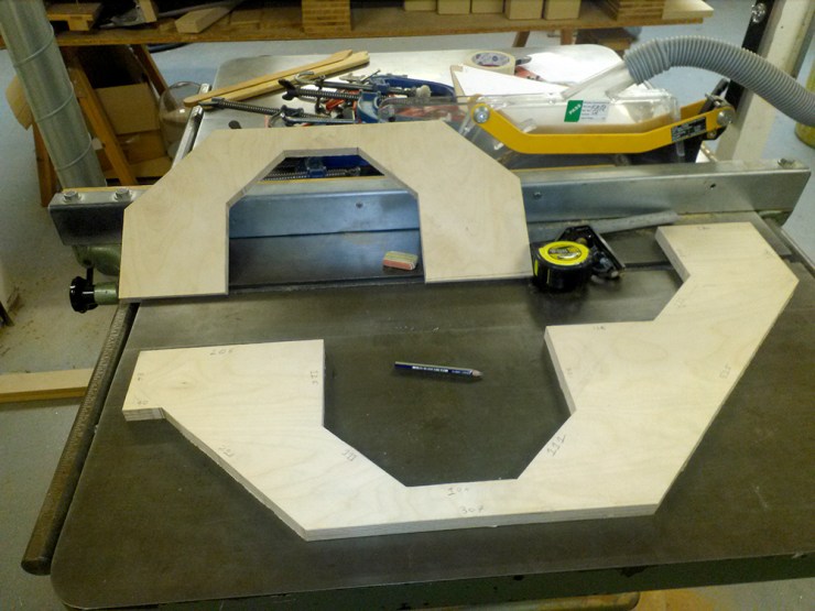
Pieces prepared in the workshop, to sit on the capitals and support the artwork

Hand-cut pieces of plywood were made to snugly fit round the capitals, so that the Dibond aluminium sheets don’t rest on the stone.

Credit: Ian Kirkpatrick

So, standing back and looking at the finished piece, looking resplendent in the winter sunshine and attracting the attention of hundreds of museum visitors, how does Ian feel?

The project was a lot of work – but it’s also been very satisfying to see it finally installed. Although the piece initially started as a comment on contemporary British settlement, it evolved into something that explored global migration throughout all of history.  Trying to find a way to tackle a theme that big, while still remaining visually coherent, is quite tricky!  But I was really pleased with the results and love seeing the finished piece housed within the magnificent neo-Gothic architecture of the Museum!


Movers and settlers

Our new exhibition Settlers, which opens today, shows that the history of the people of Britain is one of movement, migration and settlement. Here, exhibition writer Georgina Ferry finds that Britain has been receiving new arrivals since the last Ice Age.  

In Britain following the Brexit vote, the word ‘migration’ has taken on an emotional and political charge. A new exhibition opening today takes a long-view of the movement of people, looking in particular at how migration has formed the British population.

Settlers: genetics, geography and the peopling of Britain tells the story of the occupation of Britain since the end of the last Ice Age, about 11,600 years ago. From this perspective, today’s pattern of movement into and out of the country is only the latest in a long history of alternating change and stability that has made the people of Britain who they are today.

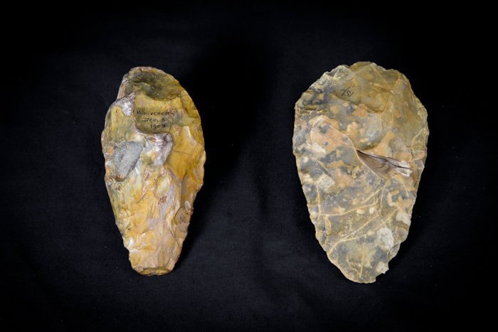
Hand axes from Wolvercote, Oxford
About 340,000 – 300,000 years ago, when conditions were slightly warmer than at present, Neanderthal hunters lived alongside a channel of the Thames near Oxford where the village of Wolvercote now stands. They made flint hand axes – all-purpose butchering, digging and chopping tools. They hunted animals now extinct in Britain.

Tracing these movements has been a fascinating detective story, with clues coming from many different types of evidence. The starting point for Settlers is a remarkable study carried out by Oxford scientists, who used DNA samples from contemporary British volunteers to trace the origins of the people who settled Britain between the end of the Ice Age and the Norman Conquest of 1066. One striking finding is that the bonds that unite Celtic communities in Cornwall, Wales and Scotland are largely cultural – genetically these groups are quite distinct.

Drinking horn finial of copper alloy and glass, 9th century – Northern Ireland. The Ashmolean Museum, University of Oxford

The genetic evidence adds a new dimension to the archaeological story, based on artefacts left behind by our ancestors, or other historical signposts such as place names. For example, although occupying Roman armies left us the names of their forts and cities, they don’t seem to have left much of their DNA. They came, saw and conquered, but didn’t stay in large enough numbers to make a genetic impact on the native British population. In contrast the Anglo-Saxons, who arrived after the Romans withdrew, left a strong genetic signature everywhere except Wales and the Scottish Highlands.

It took 2,000 volunteers and software that can distinguish tiny differences to arrive at the various regional clusters that came out of the study. When you visit the exhibition, you can play a fascinating interactive lottery game to see just how unlikely it is that genes from any specific ancestor of more than a few generations will still be in your DNA.

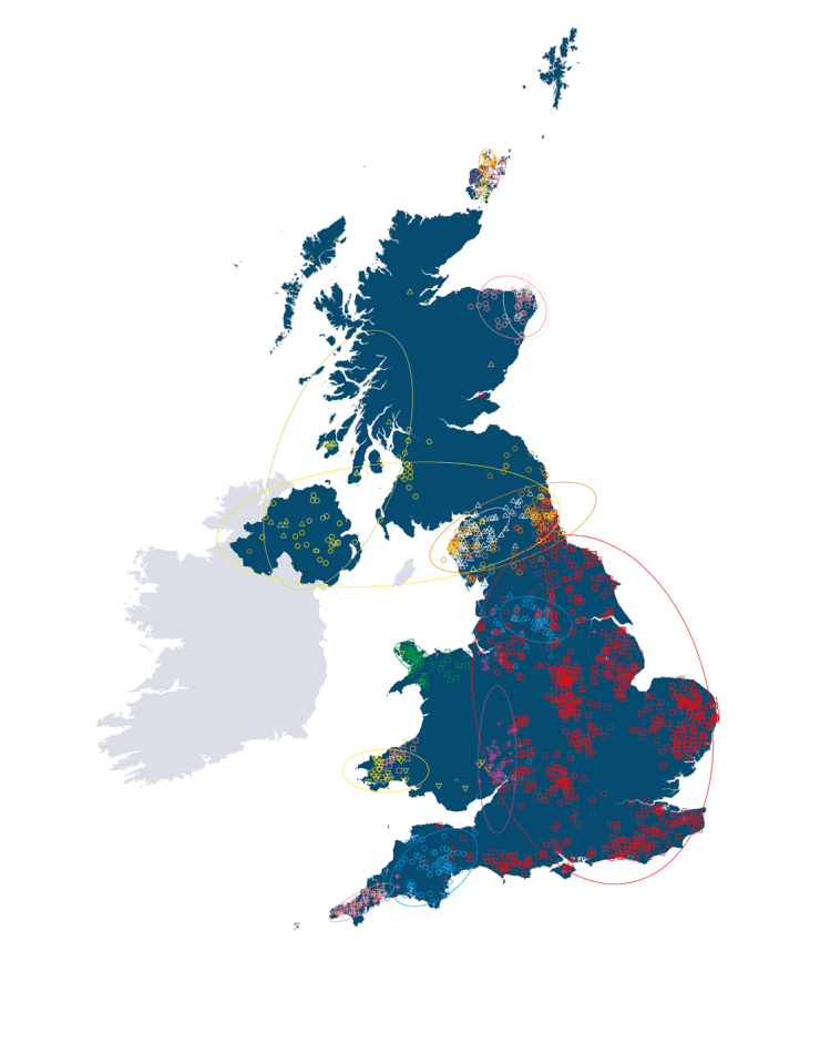
This map, created by the People of the British Isles study, is the result of comparing patterns in the DNA of a carefully selected sample of around 2,000 modern British people. It provides new evidence about links between genetic ancestry and geographical origins.

The story of movement and settlement doesn’t stop in 1066. Researchers in Oxford’s School of Geography have plotted census data since 1841 against global events, from the persecution of Russian Jews to the enlargement of the European Union, to illustrate the ebb and flow of people from and to Britain that has produced the current population mix. Another interactive lets you compare your own family’s journey with those of all the other visitors.

We will have to wait until the census of 2021 to know what a difference Brexit will make, but we can be sure that people will be arriving and leaving for a lot longer than that.

www.oum.ox.ac.uk/settlers

All that glitters…

The latest display in our changing Presenting… series showcases some of the incredible colours seen in many insects. Zoe Simmons, collections manager in our Life Collections, explains how such wonderful hues are created.

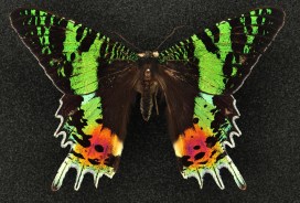
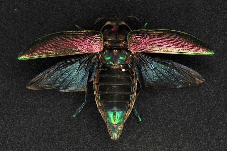
Reflected and refracted light creates the many bright and shining colours found in some insects. The dazzling natural display shown in the specimens here is formed through a combination of embedded pigments and sculpted surfaces on each insect’s external skeleton.

Some species can be variable in colour. Here a pair of Lamprima, a genus of Stag Beetles, shows off the range of colours present in the species.

Different pigment chemicals are responsible for different colours. Carotenoids produce yellow, orange and red hues, while bilins may be green, or blue if linked with proteins. They reflect and absorb different wavelengths of light, and the wavelengths that are reflected are the ones that we see as colour. Typically humans can see wavelengths of 390-700 nanometres, with the lower wavelengths perceived as blue, and the higher ones as red.

Many of the Leaf Beetles (Chrysomelidae) exhibit metallic colours.

Many insects also have multiple thin layers over their upper surfaces to help protect them and prevent dehydration. Variations in thickness and chemical composition of these layers can interfere with the transmission of light, refracting and scattering it back.

Some of the most striking metallic colours are found in the genus Chrysina, where species can be rose, silver or gold.

The shape of the surface layer can reflect light in a multitude of directions, with micro-folds, grooves, pits, hairs and scales all helping to produce complex colours and effects.

The formation and purpose of these colours is scientifically interesting, with research having applications in areas such as nanotechnology. But these insects are also simply beautiful examples of the spectacular diversity of the natural world.

Sunset moths (Uraniidae) are so called because of the dazzling array of colours on their wings. As day-flying moths they are brightly coloured like many butterfly species.

The crucial cortex

Lance Millar_Developmental Anatomy_14Oct2017.jpg-large

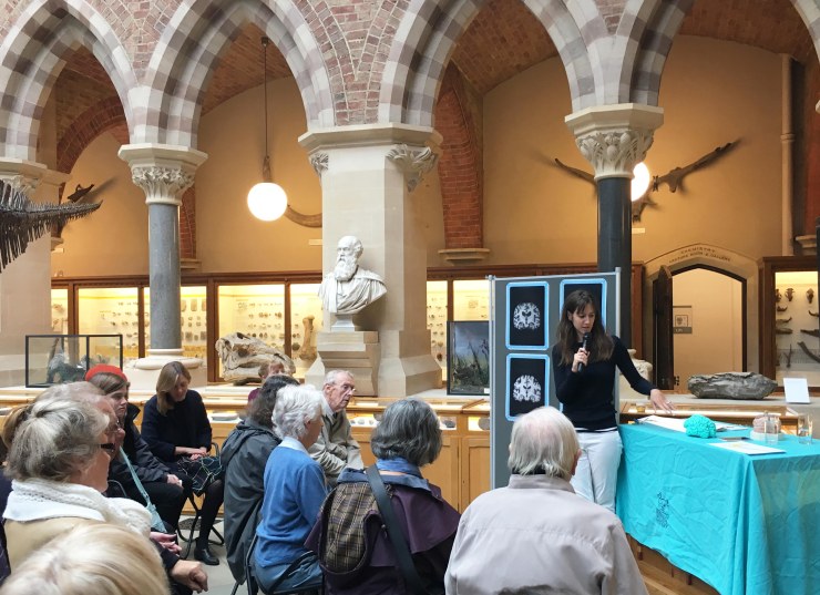
University of Oxford PhD student Lance Millar recently ran one of our Brain Spotlight events as part of the Brain Diaries exhibition programme. Here, Lance explains his research into neurodevelopmental disorders and possible treatments.

The brain has always been a fascinating organ for me. It is the site of our intelligence, our problem-solving and social skills, and it allows us to connect our senses to the world around us.

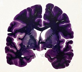
The large, folded outer part of the human brain is called the cortex, and is responsible for decision-making, language, face recognition, and a lot of the other things that I like to think are what make us human. The word cortex comes from the Greek for husk or outer shell, which underestimates the importance of what the cortex does.

Humans can survive with damage to the cortex, but depending on the part of the cortex that is damaged, a range of disabilities can result. People who have had a stroke can lose part of their cortex, leading to limb paralysis, loss of speech, or loss of memory, depending on the site of the damage.

B0009564 Human brain, coronal section, LM
Cerebral cortex – Professor Michael R Peres – Wellcome Images

Some people are also born with a developmental problem in the cortex, and are said to have a neurodevelopmental disorder. Such conditions are thought to include autism, schizophrenia, ADHD, and even dyslexia – all fairly common conditions. The damage to the cortex is subtle and complex in these conditions, and scientists are still working out exactly what happens to the brain during its prenatal development.

I am studying one particular neurodevelopmental disorder caused by lack of oxygen at birth. It is known to medical specialists as neonatal hypoxia ischaemia. The image on the right shows a cross-section MRI scan of a normal newborn human brain, alongside some babies who have been damaged by oxygen deprivation. You can see that the brains are smaller, the cortex is less folded and it takes up less space inside the skull.

Woodward
MRI scans of normal newborn brains alongside those of babies who have been damaged by oxygen deprivation. Image: Woodward et al., New England Journal of Medicine, 2006

Scientists still don’t know how to protect the newborn brain from these injuries. Some are caused by inflammation which is a normal response to illness, but can wreak havoc in the confined space of the skull. Some is caused by the presence of free radicals, which are thought to contribute to ageing and organ failure, as the newborn brain doesn’t have many antioxidants to fight these chemicals. It’s also possible that the electrical signals that neurons within the brain send to each other contribute to the damage when there isn’t enough oxygen to feed them.

So what can we do to treat oxygen deprivation at birth? One breakthrough treatment currently available is known known as hypothermia. In this technique, the baby is cooled to 33℃ which slows down the brain-damaging chemical reactions which in turn protects the brain. This is currently the only treatment available, but I am involved in the study of possible alternatives.

We don’t want to introduce any drugs to the baby’s system as they might be harmful to normal development. So scientists are currently working on treatments which help the baby’s natural body proteins to protect the brain. I do this by looking at neurons under the microscope, and identifying proteins expressed by these neurons using fluorescent probes known as antibodies.

Neurons under the microscope
An example of neurons under the microscope. Image: Lancelot Millar

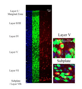
These neurons are expressing neuroserpin, a natural brain protein which decreases inflammation and cell death. I’m looking at exactly where neuroserpin is expressed in the brain, how it can be upregulated in response to oxygen deprivation, and how its chemical reactions could be used to protect the brain.

Another way to help people with neurodevelopmental disorders is to better understand how the cortex connects to other parts of the brain and how it can carry out complicated decisions. There is still so much to understand about the complexity of the human brain, and what seems like fundamental research could generate the springboard for new ideas for neurodevelopmental disorder treatments.

To explore the structure of the human brain and compare it to that of other animals see the Brain Diaries Brain Explorer below.


All about Alzheimer’s

University of Toronto research fellow Jacqueline Zimmermann recently ran one of our Brain Spotlight events as part of the Brain Diaries exhibition programme. To mark World Alzheimer’s Day today, here Jacqueline tells us about the neurophysiology of Alzheimer’s disease and the risk factors we can actively reduce to lead happier, healthier, and longer lives.

Almost all of us have in some way been affected by Alzheimer’s disease, which makes the quest for a cure that much more personal. An estimated one in nine people over the age of 65 will develop the disease, and this risk also increases with age, according to the World Alzheimer’s Report in 2015.

Brain Spotlight
Jacqueline Zimmerman’s Brain Spotlight on Saturday 16 September as part of the Brain Diaries exhibition series of events.

Due to chemical toxins, and increased longevity, the incidence for Alzheimer’s disease is on the rise. But the good news is that there is a lot that you can do to reduce your risk. At the John Radcliffe Hospital in Oxford, hundreds of scientists are currently working towards identifying the cause and the solution to the disease.

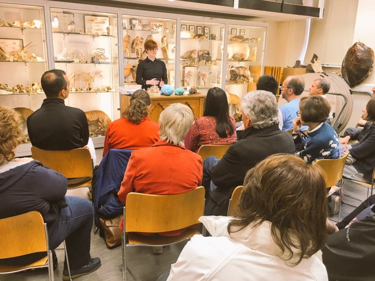
At the Brain Spotlight event at the Museum I presented images of ageing brains, and explained how brains affected by Alzheimer’s have reduced volume in the temporal lobe and the hippocampus, regions critical for language and memory respectively. Diseased brains will also often show reduced frontal lobe volume, which may reflect the changes in personality and the ability to engage in planning which area associated with Alzheimer’s. The overall volume of the brain is also reduced in sufferers because cellular changes lead to the death of neurons.

Brain Atrophy in Alzheimer’s disease
Brain Atrophy in Alzheimer’s disease. Note: overall brain volume is reduced, hippocampal regions and frontal regions are particularly affected, and ventricles are enlarged. Image: http://www.lookfordiagnosis.com

Recently, a number of genes have been identified that are related to early onset Alzheimer’s, which is quite rare and much more hereditary than late-onset Alzheimer’s. At the Nuffield Department of Clinical Neurosciences, where I am a visiting researcher, we are investigating a late-onset Alzheimer’s risk gene called Apolipoprotein 4 (APOE4), looking at how it relates to subtle cognitive impairments in middle-aged people. Working with the Oxford Biobank we are trying to determine which cognitive assessments may be most effective in predicting these impairments.

Jacqueline Zimmerman
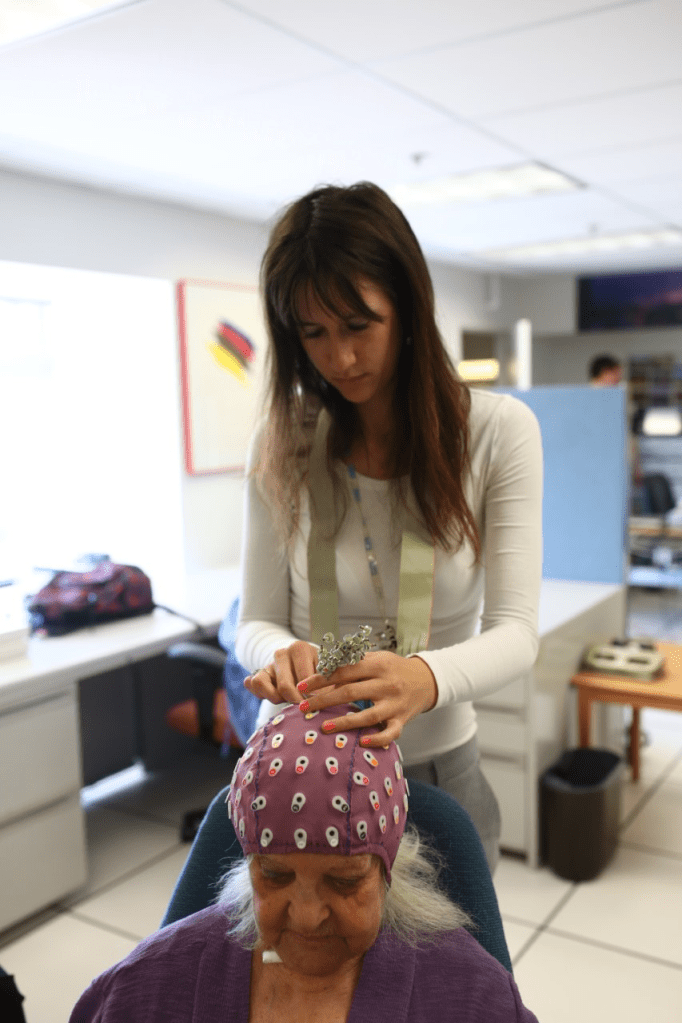
Functional brain imaging using electroencephalography at Rotman Research Institute in Toronto. Image: Rotman Research Institute

Although some of us may be more susceptible to Alzheimer’s than others, there are a number of environmental factors that contribute, including air pollution or additives in our food, like nitrogen-based chemicals which are used to preserve and flavour processed foods. It is important to reduce cholesterol in the diet, eat plenty of fruits and leafy greens, and engage in frequent physical and mental exercise.

Though there is speculation about the effectiveness of ‘brain games’ and how they translate into improvements in cognition in the real world, there are certainly large benefits of keeping your brain active.