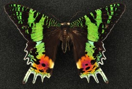
The latest display in our changing Presenting… series showcases some of the incredible colours seen in many insects. Zoe Simmons, collections manager in our Life Collections, explains how such wonderful hues are created.
Reflected and refracted light creates the many bright and shining colours found in some insects. The dazzling natural display shown in the specimens here is formed through a combination of embedded pigments and sculpted surfaces on each insect’s external skeleton.
Some species can be variable in colour. Here a pair of Lamprima, a genus of Stag Beetles, shows off the range of colours present in the species.
Different pigment chemicals are responsible for different colours. Carotenoids produce yellow, orange and red hues, while bilins may be green, or blue if linked with proteins. They reflect and absorb different wavelengths of light, and the wavelengths that are reflected are the ones that we see as colour. Typically humans can see wavelengths of 390-700 nanometres, with the lower wavelengths perceived as blue, and the higher ones as red.
Many of the Leaf Beetles (Chrysomelidae) exhibit metallic colours.
Many insects also have multiple thin layers over their upper surfaces to help protect them and prevent dehydration. Variations in thickness and chemical composition of these layers can interfere with the transmission of light, refracting and scattering it back.
Some of the most striking metallic colours are found in the genus Chrysina, where species can be rose, silver or gold.
The shape of the surface layer can reflect light in a multitude of directions, with micro-folds, grooves, pits, hairs and scales all helping to produce complex colours and effects.
The formation and purpose of these colours is scientifically interesting, with research having applications in areas such as nanotechnology. But these insects are also simply beautiful examples of the spectacular diversity of the natural world.
Sunset moths (Uraniidae)are so called because of the dazzling array of colours on their wings. As day-flying moths they are brightly coloured like many butterfly species.
The latest display in our changing Presenting… case showcases a wonderful array of dung beetles. Darren Mann, head of our Life Collections, tells us why they are so important.
Worshipped during ancient Egyptian times, dung beetles have a long history of human appreciation. Jean-Henri Fabre (1823-1915), one of the first to popularise insects in his writings, began his Souvenirs entomologiques series with the Sacred Scarab, and even Charles Darwin appreciated the weaponry adorning many dung beetles.
Dung beetles can be divided into three main groups based on their nesting behaviour. The rollers, often seen on television wildlife documentaries, make a ball out of dung and roll it some distance before burying it. The tunnellers dig directly below the dung pile and bury as much as needed for nest construction. Finally, the dwellers nest within the dung pile.
The South American Phanaeinae is one of most colourful groups of beetles. They are often referred to as Rainbow Scarabs due to their bright metallic bodies. We don’t fully understand why these beetles are quite so colourful.
Dung beetles are one of the more popular groups of insects used in ecological and evolutionary research today. They can help us to understand questions about how biodiversity loss impacts on ecosystems, or act as model organisms in the field of evolutionary development.
Unlike the much-publicised importance of bees and their pollination services, dung beetles are relatively unknown, despite their huge ecological and economic value. Their feeding and nesting behaviours provide many useful ecosystem services such as dung removal, pest fly control, parasite suppression, nutrient cycling, plant growth enhancement, improvement of soil structure, secondary seed dispersal, and a possible reduction of greenhouse gas emissions.
Through these activities, one study calculated that dung beetles are worth around £367 million a year to the UK cattle industry alone.
The largest dung beetles belong to the genus Heliocopris, which can reach up to 69 mm (pictured is Heliocopris dominus). These large beetles specialise on elephant and rhino dung. From around 2 mm in length is the oriental genus Panelus. These small beetles probably feed on the ‘dung’ of other insects and fungi.
Ancient Egyptians believed that the dung beetle kept the Sun moving across the sky like a giant ball of dung, linking the insect to the god of the rising sun Khepri. Some historians believe that it was through observing dung beetle behaviour and biology that Egyptians developed ideas about life after death.
The two most widely depicted species in Egyptian art are Kheper aegyptiorum and Scarabaeus sacer. Nowadays, only Scarabaeus occurs in this region of Africa; Kheper is now a more southern species, possibly indicating climatic changes since Ancient Egyptian civilization.
Kheper aegyptiorum on display in the museum’s Presenting… case
The UK has about 60 species of dung beetle and most of these belong to the ‘lesser dung beetle’ subfamily Aphodiinae. The largest of our dung beetles are the Dor Beetles which can reach 28 mm. Our smallest, Plagionus arenarius, is a meagre 2.5 mm. Sadly, over 50 per cent of our dung beetles are in decline due to agricultural intensification, pesticides and habitat loss.
The museum holds the only remaining soft tissue of the extinct dodo known anywhere in the world. The partially dissected skin of the head and scales from the feet of a single dodo represent one of natural history’s most iconic specimens. In fact, it is so tied to the museum’s identity and history that we use the dodo as our logo and it is even incorporated in the name of this blog.
Although the dodo head had been at Oxford University since the formation of the original Ashmolean Museum in the 17th century, it wasn’t really until the 19th century that the specimen really became celebrated.
Around this time, publications confirmed the extinction of the dodo from the island of Mauritius, where it was endemic. To capitalise on the rising interest in the animal, Ashmolean Museum Keeper John Duncan commissioned a number of casts of the Oxford Dodo head to give to, and exchange with, other museums.
One of the earliest of these casts was presented to the British Museum in 1828; later casts are recorded as being sent or exchanged with leading scientists of the time, as well as with Leiden Museum and the Royal College of Surgeons.
From these original and later casts further casts and models were presumably made, and eventually, dodo specimens spread to virtually every major natural history museum in the world. Today, many museums display casts of this head, all stemming from the single specimen held here in Oxford.
One of the many casts in the Oxford University Museum of Natural History, this one has been painted to match the original specimen
The Museum contains a number of models and casts of the head too; some are made from plaster and resin, some are painted to resemble the original specimen. The head of the dodo was actually dissected in 1847, by Henry Acland. He removed the skin from one side of the face so the early casts are a record of how the specimen would have looked originally.
In preparation for the Presenting display in the Museum I contacted natural history museums through the Natural Sciences Collections Association asking people to share information and photos about their casts and models of the dodo head. I wanted to try and construct a picture of how the dodo head was disseminated, as well as capture the diversity of quality and colours of representations of the original specimen. Here’s how far some of the dodos have flown:
Cast of the head labelled as coming from Cartwright Hall. Curator Gerry McGowan suspects this may have come via the Bradford Philosophical Society collections. The first curator of the society, Louis Compton Miall was friends with Thomas Henry Huxley and through him had contacts with many other geologists who may have gifted or exchanged this cast.
Bristol Museum & Art Gallery Bristol received a cast of the head directly from Oxford from Philip Duncan in 1834, keeper of the Ashmolean Museum between 1826 and 1855. Unfortunately, the head was likely destroyed in bombings of Bristol in 1940.
Canterbury Museum, New Zealand
Received a cast of a head from Professor Rolleston on 21 July 1871 in exchange for two kiwi skeletons which are still in the museum collections today.
Cast of head and foot presented to the museum by E.Ray Lankester in 1891/1892 just after leaving UCL and being appointed the Linacre Professor of Comparative Anatomy at Oxford.
The Great North Museum Hancock’s cast was presented by George Townsend Fox. This specimen had been presented to the Natural History Society of Newcastle in 1841 by Fox and had originally been presented by Philip Duncan.
Cast of the head that had quite a circuitous route to the Horniman Museum. The Horniman received the cast from the geology department of Queen Mary’s University of London in 1964 which received the cast from the Saffron Walden Museum in 1962.
Manchester Museum Cast of a head, presumed to have been presented by William Boyd Dawkins. The cast is currently on display in the Living Worlds gallery in Manchester Museum.
The National Geological Repository British Geological Survey
The Natural History Museum’s collections contain three casts of the Oxford Dodo head – two in the ornithology collections (pictured) and one in the palaeontology collections. The unpainted cast in the ornithology collections has the name ‘Johnson’ inscribed into the base.
Colleague Adam Smith got in touch with some interesting specimens from Wollaton Hall. The first one looks like another cast in this series but the second cast is unlike any of the others gathered here. The cast shows an open eye, detail on the beak as well as a more defined hook to the end of the bill.
Unfortunately, there’s not much information about the origins of these two casts so it’s probable that the ‘open eye’ cast may be a cast of a model reconstruction or an in progress sculpt. There’s an extremely slight chance it’s a cast of an otherwise unknown dodo head… If you recognise this dodo head do get in touch so we can solve this mystery for colleagues in Nottingham (it’s not the model dodo that we have on display here!).
Cast of a head at Warwickshire Museum with damage to the beak. Donated to the museum by clergyman and naturalist Reverand Andrew Bloxham in the 19th century. As the museum is currently moving stores, further information about when this cast was acquired is inaccessible.
**
If you work at a museum and have a dodo head cast to share, please do get in touch and we’ll update this blog accordingly.
Last updated: 10/11/17
‘Presenting… The Flight of the Dodo’ was on display at the Museum of Natural History from the 25 January to 22 March 2017.
Acknowledgements
With many thanks to colleagues across the sector who helped with information and images about dodo specimens: Adam Smith, Alice Adams, Jack Ashby, Carol Davies, Bonnie Griffin, Dan Gordon, Yvette Harvey, Mike Howe, Emma-Lousie Nicholls, Laura McCoy, Gerry McGowan, Nigel Monaghan, Henry McGhie, Pat Morris, Paul Scofield, Paul Shepherd, and Paul Sweet.
The latest display in our changing Presenting… case showcases some pioneers of photography and features a recently-discovered original print of Charles Darwin by Julia Margaret Cameron…
In the early days of photography, in the mid-19th century, a number of photographic innovators had close links to the Museum and its collections. Perhaps the most famous of these is Charles Lutwidge Dodgson (1832-1898), better known as Lewis Carroll. Dodgson was an author and mathematician at Christ Church College in Oxford, and also an accomplished early photographer.
Dodgson’s friends were often the subjects of his photos. A well-known example is Alice Liddell who, as well as sitting for portraiture, provided inspiration for Dodgson’s book Alice’s Adventures in Wonderland, as did the Dodo in the Museum’s collections.
The image at the top of the article is titled The Anatomy Lesson with Dr George Rolleston and was taken by Dodgson around 1857. It shows Dr Rolleston, Professor of Medicine at the University of Oxford at the time, pictured with a selection of specimens from the Museum’s collections.
Two of Dodgson’s subjects were accomplished photographers in their own right, and perhaps the most famous female photographers of their time: Julia Margaret Cameron (1815-1879) and Sarah Angelina Acland (1849-1930).
Cameron’s innovative and distinctive style of portraiture raised photography to a true art form. Her intentional use of shifted focus and experiments with process and finish gave an ethereal look to her works resembling the artistic style of the contemporary Pre-Raphaelites.
Many prominent scientists sat for Cameron, including naturalist Charles Darwin. She took the portrait above in 1868 at her home on the Isle of Wight. An original print of this photograph, made by Cameron, was recently discovered in the Museum Archive, uncatalogued. It features her signature on the back and the blind stamp from her printseller in London on the front.
‘Mr Ruskin & his old friend at Brantwood’ by Sarah Angelina Acland, 1893
Sarah Angelina Acland, inspired and taught by Cameron, is best known for her innovations in colour photography, but she also took many black and white photographs while learning the art form. The portrait above features her father Sir Henry Wentworth Acland and the artist John Ruskin at his home. Both Ruskin and Acland were instrumental in the founding of this Museum, and Sarah even helped to lay the founding stone in 1855.
In a 1904 Royal Photographic Society exhibition Acland was the first to exhibit work combining images taken with red, green and blue filters, three years before the launch of the colour photography process invented by the Lumière brothers.
These images, including the original print of Charles Darwin by Julia Margaret Cameron, are on display in our Presenting… case until 24 January.
by Juliet Hay, Earth Collections preparator and conservator
I feel myself very lucky to have a job that involves working with the fossil remains of long-extinct animals. One of the things my colleagues and I are currently working on is a plesiosaur – a marine reptile that lived in the sea millions of years ago.
This particular specimen was found in a clay pit near Peterborough by members of the Oxford Clay Working Group in 2014, and is a near-complete example of its kind. The palaeontologists who found the specimen named it Eve, although we don’t know if it was male or female, and perhaps never will.
The discovery of large fossil vertebrates like this is rare, so we are fortunate to have had the specimen donated to the Museum by the quarry owners Forterra.
Juliet at work on the plesiosaur skull
The plesiosaur is 165 million years old and, when alive, was around 5.5 metres long. It had a long neck, a barrel-shaped body, four flippers and a short tail. The find is particularly exciting as the skull was also discovered. It is encased in a clay matrix, which is relatively easy to remove, but the work has to be carried out under magnifying lenses and microscopes.
As the skull is quite small relative to the size of the body, the features are very delicate and it is a painstaking process to remove the sediment without damaging the fossil bone or losing any tiny fragments. Fortunately, pictures of the skull have been produced using CT scanning technology, and the images are proving invaluable as an aid to assist in its preparation. It’s a bit like having a jigsaw puzzle with the picture on the lid to refer to!
A belemnite hooklet at 12x magnification, found with the plesiosaur remains and possibly part of Eve’s last meal
The clay covering the skull is being sieved and examined and tiny hook-shaped fossils have been found. These came from the arms of squid-like creatures called belemnites, which may have formed a large part of the plesiosaur’s diet.
It is too early to say for sure, but Eve could represent a species new to science, as some features, such as the shape of the flipper bones and some of the surfaces of the bone in the skull, are quite unusual. Further research needs to be done before the findings can be published in scientific journals – watch this space.
And if you’re visiting the Museum before 25 July, you can see some of the fossilised remains of Eve for yourself, in our Presenting… display case.
Since the launch of our Kurt Jackson exhibition in March, the Museum has gone a bit bee mad. We’ve had themed events for all different audiences; expert beekeepers sharing their top tips with adults, right through to little ones trying on beekeeping outfits and building a giant beehive. There’s even more coming soon, including a talk by Kurt, the artist himself, on 12th May.
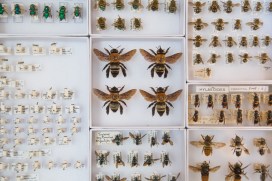
Now the buzz has spread to the Presenting… case, our changing display that shows off real treasures from the collection. The Jackson exhibition focuses on some of the 270 British bees, but this smaller display gives a different perspective, exploring the amazing variety of bees found around the world.
A selection of specimens from the Museum’s collection, which shows how varied bees can be.
Bees are one of the major groups of insects, numbering about 20,000 described species. Only a very small proportion of bees are the familiar honeybees or bumblebees that we think of first – most species are actually solitary bees. Bees exist in a great diversity of shapes, sizes and colours. In particular the smaller species do not look like bees at all and are often mistaken for small wasps or flies.
Many bees are specialised as pollinators and have evolved together with flowering plants for over 100 million years. In return for pollination services, plants provide nectar, pollen and other substances to bees.
Although most are specialist pollinators, about 10 per cent of bee species are parasites of other bees, taking advantage of the nectar and pollen collected by their host to feed their own young. These parasitic bees can be quite strange in appearance – not needing to collect pollen they have typically lost most of their hair and appear more like wasps.
The Museum has one of the most important bee collections in the world, containing specimens collected over 200 years ago and from many different countries. The star bee specimen, and one of the Museum’s greatest treasures, has to to be Wallace’s Giant Bee (Megachile pluto). This is the first time it has been on open display to the public.
Wallace’s Giant Bee (Megachile pluto) alongside a honeybee (Apis mellifera)
It was captured by the Victorian explorer and naturalist Alfred Russel Wallace in 1859. Only found on the Indonesian island of Bacan and its two neighbouring islands, this giant was believed to be extinct until it was re-discovered in 1981. The massively enlarged mandibles of the female are used for collecting tree resin and excavating tunnels in termite nests. To give an impression of scale, Wallace’s Giant Bee is shown here next to a familiarhoneybee (Apis mellifera).
You have just a couple of weeks left to appreciate the wonderful diversity of bees, before it closes on 16th May.


 Although most are specialist pollinators, about 10 per cent of bee species are parasites of other bees, taking advantage of the nectar and pollen collected by their host to feed their own young. These parasitic bees can be quite strange in appearance – not needing to collect pollen they have typically lost most of their hair and appear more like wasps.
