What a year…
At the end of 2013, the Museum closed for a 14-month roof refurbishment project. On 15 February 2014 – a year ago yesterday – we reopened and returned to the light. Here’s what happened…

Oxford University Museum of Natural History Blog

At the end of 2013, the Museum closed for a 14-month roof refurbishment project. On 15 February 2014 – a year ago yesterday – we reopened and returned to the light. Here’s what happened…

Amo Spooner from the Museum’s Life Collections has been out in the Museum sharing some of her favourite objects. Here’s the latest in our Spotlight Specimens series…
**

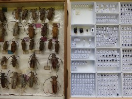
Big impressive beetles or small shiny ones? That is the question. For me it’s all about the small ones, but here I am getting people’s (and the T. rex’s) attention with the big ones. It’s my tactic for engaging their interest before I try to convince them that the small ones are so much cooler!
Monday – Thursday at 2.30pm a member of the Museum’s collections staff can be found out in the Museum talking about something interesting. For my latest session of Spotlight Specimens I chose to show off drawers of my favourite beetles.
 The big ones are from a family of beetles called Cerambycidae or Longhorn Beetles. This family is found all over the world and varies greatly in size and colour. These ones are particularly interesting to me because of the historic collection they are from. The vast Baden-Sommer collection, containing many different beetle families, came to the museum via a dealer in 1910 and unusually it is still in its original layout. The labels you can see in the drawer were written by the two entomologists that collected the specimens, J. Baden and M. Sommer.
The one you see in my hand (above) is in the subfamily Lamiinae – also charmingly known as Flat Faced Longhorns.
Part of my job is to re-curate and move historic specimens into pest-proof housing – I am currently writing a blog post explaining this, so watch this space! In a nutshell, the Baden-Sommer Longhorns are a good example of drawers in need of some TLC. This leads me nicely on to my second choice of drawer, the Histeridae.
 These are my first love when it comes to beetles. The Histeridae, or Clown Beetles, vary a lot in size; the one in my hand (below) is about as big as they get, but they can be as small as 1 mm in length.
 I have re-curated all of the Museum’s historic Histeridae specimens and mounted up many modern ones, like you can see above. This modern system of trays and pest proof drawers ensures the longevity of specimens, as well as making them easier to access.
So what makes the little ones so special? During the afternoon I met visitors from home and abroad, young and old. I convinced them to to look a little closer, admiring their shiny black armour and fascinating adaptations. I think they finally agreed that big isn’t always best.
Amo Spooner, Collections assistant (Life)
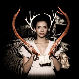

I’ve just received some fabulous pictures from a photo shoot here in the Museum. The building and its specimens are shown at their very best and the model’s striking looks add a sheen of glamour to each photo. But what makes these images really special is that the model is one of our own; Aisling Serrant is better known to Museum staff as a trainee education officer on the HLF Skills for the Future programme.

Aisling spent four months in the Public Engagement team in 2014, largely working with school groups and families. Not always the most glamorous job. In November 2014 we had the opportunity to see her in a completely new light. Oxford Fashion Week was coming to the Museum and they were running open casting sessions for models. Aisling remembers how she got involved:
**
When the initial meetings started taking place between the Fashion Week organisers and the Museum staff, my ears pricked up. Alongside studying or working I have modelled for years. My degree is in archaeology, so people have always found the combination with fashion modelling quite funny – perhaps imagining me standing knee-deep in a muddy trench in stilettos! It seemed too good to be true – could there really be a chance for me to bring my two contrasting types of work together?

I was delighted when I was asked to model in all three shows that were to be held at the Museum. They took place on Friday 7 and Thursday 8 November.
Friday night was a busy one with two shows in one night. I felt right at home, with the familiar faces of old friends like the T.rex and Iguanodon (oh and some of the staff members too!). However it did all feel a bit surreal.

The Museum Annexe had been transformed into the backstage area, but the last time I had spent so much time there I’d been running an archaeological dig activity with year 6 children in the Making Museums project. Now the place couldn’t have looked more different with rails upon rails of clothes, photographers’ flashes and the distinct smell of hairspray in the air.
Saturday night was the big finale to the week – the Birds of Paradise show in the Museum central court. The skeleton parade was parted so we could walk down the middle and the Triceratops skull was moved to become the backdrop for the first part of our walk. The museum was transformed into another world for the evening.

The addition of atmospheric music and such stunning outfits was truly breathtaking – enchanting at times, slightly eerie at others – but always fantastically dramatic. The nature-inspired outfits, some smothered in black feathers, others twinkling with jewel beetle shells, served as a reminder of how extraordinarily beautiful the natural world is.
**

Photographer Julia Cleaver was here for the shows and was so inspired by the venue and so enjoyed working with Aisling that she returned recently to do an extra photo shoot. These photos are the stunning outcome of that session. Many thanks to Julia for letting us share them here.
Rachel Parle, Interpretation and Education Officer

As a Museum research fellow, my work on arthropod palaeontology often takes me to exotic places to examine and collect fossils. I recently returned from a packed five-week trip to Australia and Argentina. During this time I managed to squeeze in two fieldwork trips, a museum visit to examine some collections, and an international conference.
It began in early September when I flew to Adelaide, Australia, to meet up with friends and colleagues at the South Australia Museum (SAM) for some fossil-collecting fieldwork. A group of eight of us piled into fully loaded trucks and started the drive to Cape Jervis, where we boarded the ferry to Kangaroo Island. On this beautiful island, there is a spectacular fossil site known as the Emu Bay Shale. The fossils here preserve 510 million year old Early Cambrian animals in incredible detail, including soft parts not normally found in fossils, such as eyes, gills, skin and guts.

Dr John Paterson, Dr Diego Garcia-Bellido and other researchers from the SAM have published numerous papers on the weird and wonderful animals from this site. I had already been fortunate enough to work with these guys on the anomalocaridids – very early marine animals – from the Emu Bay Shale a couple of years ago. After the fieldwork this time, we returned to Adelaide with a truckload of fossils to add to the SAM collections. I then spent two weeks working in the museum on previously collected specimens, and making research plans for the years to come as part of the ongoing collaborations between this Museum and the SAM.

One of my favourite things about working in Australia is the chance for close encounters with the local wildlife, and this trip did not disappoint. During our time on Kangaroo Island, we saw many wallabies, Little Penguins, countless types of birds, and kangaroos of course. I even got to hold an echidna.
John, Diego and I then met up in Sydney airport for the long journey to Mendoza, Argentina where we joined nearly 900 colleagues for the 4th International Palaeontological Congress. This is one of the biggest conferences in our field, and takes place only every four years. We enjoyed a week of fantastic talks, including some given by the Museum’s researchers Dr David Legg and Prof Derek Siveter.
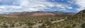
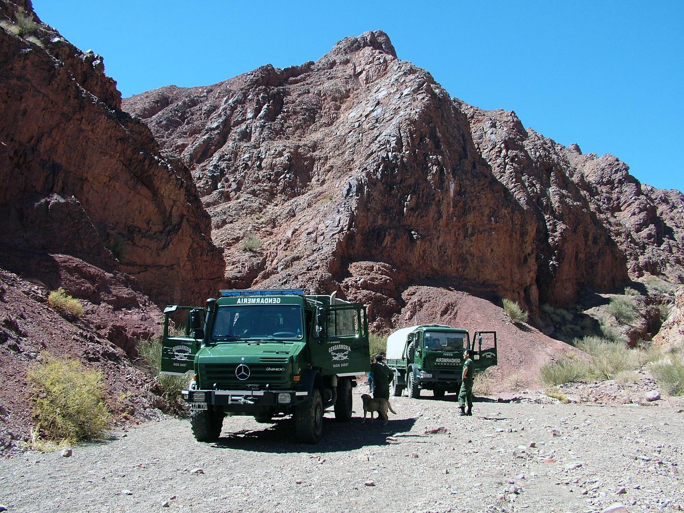
After the five-day conference, 30 of us headed out on a related field trip to the Argentinian Precordillera for a Palaeozoic marine journey to explore the wonderful rocks and fossils of western Argentina, near the border with Chile. We saw lots of lovely fossils, including trilobites, brachiopods, bivalves, corals and sponges. The terrain was so rugged at times that the field trip leaders had brought in the Argentine National Gendarmerie to transport us in army vehicles!
The scenery was spectacular, with impressive views over the Andes mountain range. After four marvelous field trip days, I then returned to Oxford, completing my journey around the world. The conversations and feedback from the conference and fieldtrip will help with my future research directions at the Museum. The fossil work in Australia provided important comparisons for the research I do here in Oxford on local collections, and will undoubtedly be the subject of future publications (and, of course, blog posts…).
Allie Daley, Museum Research Fellow
 There are many fascinating displays in the Museum, but there’s something special about meeting an expert and chatting to them about the collections they love. Every Monday to Thursday our Spotlight Specimens series gives you the chance to do exactly that.
Taking place under the T. rex in the Main Court at 2.30pm each day, staff from across the collections choose favourite specimens to share with the public. These experts will also be writing a series of Spotlight Specimens blog posts for those of you who can’t make it to the Museum to meet them in the flesh. In this, the first in the series, Gina Allnatt kicks us off with a Halloween special…
**
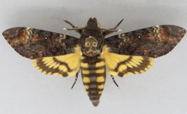
It’s October, month of falling leaves and trick-or-treating, so what better way to get into an autumnal mood than to talk about two moths with marvellously morbid names?
What do the Death’s Head Hawkmoth (Acherontia styx) and Black Witch moth (Ascalapha odorata) have in common? They are both associated with the film and novel Silence of the Lambs. The Death’s Head was used in the film, but the moth in the novel was originally the Black Witch. The moth was changed for the film for two reasons: The producers thought that a moth with a skull on its back would look more sinister, and also because it was almost impossible to get live specimens of the Black Witch moth for filming.

The author of Silence of the Lambs, Thomas Harris, may have chosen the Black Witch moth because of the many legends and myths that surround it. In Jamaica it is known as the “Duppy Bat.” In Central and South America it’s known as “Mariposa de la Muerte”, which translates as “Butterfly of the Dead” because there is a myth which claims the moth is a harbinger of death. A less sinister version of this myth suggests that if you find one of these moths in your home it means an ancestor or loved one who recently passed away is paying you a visit.
However, the subtlety of these myths would probably not translate so well on film, so Mr. Death’s Head Hawkmoth took centre stage. The vernacular name of this moth comes from the skull-like markings on its back. There are actually three species of Death’s Head Hawkmoth- A. atropos, A. styx and A. lachesis. Though the moth mentioned in the film is Acherontia styx, Acherontia atropos was actually used instead.
All Acherontia supplement their diet by raiding the hives of bees for honey. The moths achieve this by using their extremely thick cuticle, which makes them impervious to stings. But the moth also uses another tactic: it is able to emit an odour that is chemically identical to the worker bees’ scent. This fools the bees into thinking the moth is one of their own. They also emit squeaking noises while in the hive. Some scientists posit that the squeak is similar to the noise a queen honeybee emits when she wants the workers to freeze. No one has been able to observe this theory, however.

Despite all the myths and legends surrounding the Death’s Head Hawkmoth and Black Witch moth, both are large and harmless species. It is perhaps the fact that most moths are nocturnal which gives rise to so many legends and misinformation about them. It’s often the case that people will love butterflies but don’t like moths. Moths evolved before butterflies, and it is likely that the butterflies people hold dear evolved from day-flying moths (many day-flying moths exist today and are even more colourful than their butterfly counterparts!).
So remember this when you next see a moth (the original butterfly!) fluttering near a lamp as the sun slowly disappears.
Gina Allnatt, Curatorial assistant (Lepidoptera)
Gina will be talking about the Death’s Head Hawkmoth and Black Witch moth at 2.30pm on 28 and 31 October as part of our Spotlight Specimens series, running Monday to Thursday at 2.30pm.


They say a picture is worth a thousand words, and at the Museum we make thousands of pictures: pictures to document, pictures to investigate, and pictures to wow. We use a lot of different imaging techniques too, from standard close-up photography to scanning electron microscopy, which reveals the most minute details.
To coincide with the final week of the Wildlife Photographer of the Year exhibition here, on Saturday 20 September we held a new adult workshop to give people some hands-on experience of some of these processes. Imaging Techniques in Modern Natural History gave participants the chance to get up close to some wonderful specimens and make their own images to take home.
I had planned to review the day here, but Rose Parkin, who took part in the workshops, very helpfully sent in her own write-up of the sessions. So here’s a special guest post from Rose, along with some pictures taken by people on the day.
*
By Rose Parkin
When I signed up for the digital imaging course I expected a fairly dry, tech-heavy day. Instead, the experience was really exciting. Not only did it provide hands-on experience of viewing and recording images with new technology, it also gave me a brief glimpse behind the scenes of my favourite museum.
Laser Scanning and Digital Modelling
For our first session our small group was led through a maze of corridors by Sarah Joomun, the Documentation Officer, to the laser scanning lab. It sounded a bit futuristic, and it turned out that it looks that way too. Sarah popped a fossil onto a mount, clicked a few buttons and red lasers appeared, scanning the fossil’s surface while it rotated. After ten minutes the first 3D image of the fossil was produced – a beautiful net of triangles, which looked like a teleporting object in a science fiction film.

Sarah turned the fossil and scanned it again. The challenge was then to fit these two images together to make a complete 3D model. Amazingly, this technique enables other palaeontologists around the world to see and replicate, with the use of a 3D printer, the exact size and shape of a fossil without it ever leaving the museum.
Multi-plane Microscope Photography
Our next session was upstairs, with artist-in-residence and photographer Katherine Child. Even though we were close to the main corridor of the museum it felt like a real working space, crammed full of equipment and insect specimens. Katherine had chosen the tiniest of insects for us to photograph with the multi-plane microscope. It looked like a small seed with some barely visible limb-like protrusions.

But under the microscope a wonderfully strange insect became visible, with the most bizarre appendages and bright orange legs. While the microscope already showed a great deal of detail the multi-plane photography captured an incredibly crisp image. The microscope takes large numbers of photos of the specimen, using different focal planes each time, then the focussed elements are all stacked together to produce a crystal clear photograph.
Once we’d chosen and photographed some other insects from the collection and poked around the room a bit (finding a disturbing collection of large pickled spiders), we were taken on a tour of the entomology department. Katherine led us through corridors of offices and labs, up to a stunning store room that felt almost church-like, with rows and rows of cabinets full of fascinating insects.
Scanning Electron Microscopy
After lunch we had a laboratory session with museum director Paul Smith to look at sand under an electron microscope. Luckily, that was much more exciting than it sounds! The sand was taken from Dog’s Bay on the west coast of Ireland and is rich with a wide range of tiny fossilized organisms. Paul showed us how to carefully select individual microfossils from a tray using just a microscope and a paint brush.

We then viewed some of the microfossils using a scanning electron microscope. This allowed us to see an incredible level of detail. The microscope was so powerful that we could see hair holes in a fossil the size of a grain of sand.
DSLR Macrophotography
My final session was a crash course in macrophotography. Held in the seminar room, the low lighting and floor-to-ceiling collection of specimens lent an almost eerie feeling to the set-up.


Once prepped, we were let loose on four separate camera setups. Being able to choose and shoot at our own pace made this feel like a really creative experience. The help given by professional photographer Keith Barnes and public engagement officer Scott Billings was perfect – very hands on but not patronizing (despite my lack of DSLR experience).
With this digital imaging course the museum has created a really exciting snapshot of the work that goes on behind the scenes, reinforcing the fact that this impressive place is much more than just an ordinary museum.
