When the campaign to build the Museum was launched, science at Oxford was understood as natural theology. By the time the Museum opened in 1860, a new secular approach to science was on the rise.
In this last episode of the Temple of Science podcast series we see how the art and science of the Museum responded to the challenge posed by Charles Darwin’s theories of evolution and natural selection, and the scientific naturalism that they epitomised.
The Museum was not originally simply a museum as we understand it today: It was an entire science faculty. In episode four of the Temple of Science podcast series we see how the museum’s overarching principle of design – that art should be used to teach science and to inspire generations of scientists – was put into practice in some of its less familiar but no less beautiful spaces.
The central court of the Museum was described by one founder as ‘the sanctuary of the Temple of Science’. In the third episode of the Temple of Science podcast series we see how every detail of this unique space was carefully planned and crafted to form a comprehensive model of natural science.
In the second episode of the Temple of Science podcast series we take a closer look at the decoration on the outside of the Museum building.
From the outset, Oxford University Museum wanted to teach the principles of natural history through art as well as science. The carvings around the windows of the façade, incorporating designs by John Ruskin and carved by the brilliant Irish stonemason and sculptor James O’Shea, revel in the vitality of nature, while the decorations round the main entrance remind us that, for the scientists in Victorian Oxford, natural history was the study of God’s creation.
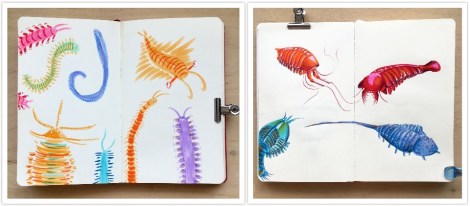
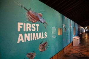
Worms, fish and … Greenland? Hugely different topics which all have one thing in common – the Museum’s First Animals exhibition online lecture series. Running every other Wednesday from May until September 2020, this series provided a fantastic insight into a wide range of topics about how the first animals lived, died, and are studied. And illustrator Rachel Simpson tells us how she drew her way through them all…
I came across this lecture series just before the first talk and I knew I had to sign up. Drawing along to lectures is a hobby I seem to have developed in the past few months as we went into lockdown and didn’t have much to do. It’s the perfect combination for me – an opportunity to listen to interesting topics and brush up on my live drawing skills at the same time. There’s no pause button, there’s no asking the webinar speaker to just go back a few slides and hold on a minute whilst I draw; it’s fast paced, it’s inspiring and it’s a great way to just create art.
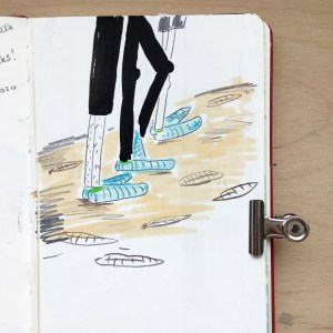
Barma Booties used on the rocks at Mistaken Point, and my first drawing of the series.
I’ve done some illustration work with the Museum before so I knew that it was going to be fun. In 2018, I worked with Dr Jack Matthews illustrating Ediacaran Fossils as part of a collaborative university project between the University of Plymouth and the Museum. I was also lucky enough to be able to go to Newfoundland and see some of the fossils myself, again with Jack. This was such an incredible opportunity and opened up a whole new world of science/art collaborative work which I didn’t know about before.
The First Animals series kicked off with Jack’s talk titled Don’t walk on the rocks! – an interesting insight into how protective “Barma Booties” (some rather funky socks worn to protect fossil sites such as Mistaken Point, Newfoundland) might actually be damaging to the fossils they’re meant to be protecting. Having been to Mistaken Point myself and worn these socks, it was interesting to hear about their possible impact and to learn about the experiments conducted to prove this fact.
Of course, at the same time as Jack was talking, I was scribbling away in my sketchbook trying to form some sort of visual response to the talk. At the end of the hour I’d managed a portrait of Jack and a family of Barma-Booted tourists trampling on the fossil site. It was a start. The beginning of my lecture drawings and a point at which I can retrospectively say started a new hobby.
Annelid worms drawn with Tombow brush pens.
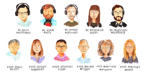
Over the following weeks we heard about worms from Dr Luke Parry; 3D reconstruction from Dr Imran Rahman; The Chronicles of Charnia by Dr Frankie Dunn; and the first animal skeletons from Dr Duncan Murdock. Luckily for me, all the speakers kindly included photos and descriptions of the topics they were discussing which meant that I was never short of visual inspiration for my drawings. After all, it’s hard to try and draw an annelid worm if you’ve never seen one before.
I love to look at the fossils being discussed and then try to draw a little character or creature inspired by them. They’re not scientifically accurate, nor are they always anatomically correct, but they have character and begin to bring to life the essence of something that’s been dead for many millennia. The fossils are obviously stone-coloured so I take as many liberties as possible when it comes to colour. I like to make them as vibrant and colourful as I can, so although they probably didn’t look like that, that’s how I like to think they looked.
Some fun little beasties from Dr. Imran Rahman’s talk.
Charnias galore! They come in all different shapes and sizes.
Small filaments which could have joined all those Charnia together.
Shells, bones and teeth from Dr. Duncan Murdock’s talk drawn in Tombow brush pen and Posca Pen.
Within my wider practice I like to use stamps as the basis of my illustrations. These however, are time consuming to make and therefore not very suitable for when I’m drawing along to lectures. As a result I’ve found myself using brush pens and pencils to make my lecture illustrations. If you’re interested in art, or thinking about getting into art, brush pens will be your best purchase. They create a wonderful quality of line and are quick and easy to use. Whereas a ballpoint pen will give you one line of a certain weight and thickness, brush pens are versatile and depending on the pressure applied, the line quality will change.
For the first few lectures I only used brush pens, but later on I decided to use coloured pencils as well, to add depth to the drawings. As I got more used to drawing in lectures I found that I was making more illustrations per talk. Early on, I managed to finish maybe a double page in my sketchbook but towards the end of the series I was filling four double pages! It’s amazing what a little bit of practice can do.
As the weeks went by the talks continued and we heard about the evolutionary origin of animals from Museum director Professor Paul Smith; an introduction to taphonomy, the study of fossilisation, by Professor Sarah Gabbott; and how the first animals moved by Professor Shuhai Xiao.
During this time I became a lot more confident drawing the specimens; looking back I can see that this was the period in which my work developed the most. My drawings began to have more character and life. The landscape drawings were slowly becoming more realistic and detailed. This was great news for me as this whole endeavour began as a way to practice my drawing skills in a timed environment.
Paul Smith’s lecture has to be my favourite of them all. He gave a wonderful talk all about the Evolutionary Origin of Animals and talked us through his fieldwork expedition to Greenland. How I would have loved to have been on that trip!
How I would have loved to have been on this trip! Drawings of Professor Paul Smith’s fieldwork to Greenland.
Some of the weird and wonderful fossils Professor Paul Smith found on his trip.
One of my favourite drawing from the lecture series! Drawn with Tombow brush pens and Polychromo pencils.
It was during Paul’s talk that I made one of my favourite drawings from the series – the plane –and coincidentally it was also at this point that I bought myself some new polychromo pencils. I started using these pencils in my illustrations on top of the Tombow brush pens. The pencils added a softer layer on top of the solid base colour from the brush pens and meant that I could add more details, shading and most importantly, the characterful eyes I love to add to my drawings.
Fish and animal studies from Professor Sarah Gabbott’s introduction to taphonomy, the study of the processes of fossilisation.
Imagine being the owner of this house and being told there were found fossils on your roof! Drawing from Professor Shuhai Xiao’s talk.
Buoyed by this development in my drawings, and some lovely responses to my work on Instagram and Twitter, I raced through the next few weeks of talks and made twelve pages of drawings over the next four talks. Professor Derek Briggs told us all about extraordinary soft-bodied fossils; Professor Gabriela Mángano told us about the trace fossil record; and Professor Rachel Wood gave us her thoughts about what triggered the Cambrian Explosion.
Another favourite drawings from the series, drawn from Professor Derek Briggs’ talk.
Close up of drawing from Professor Derek Briggs’ talk.
Trace fossil studies drawn in Tombow brush pens and Polychromo pencils.
The last drawings from the series from Professor Rachel Wood’s talk.
Another of my favourite drawings from the series was from Derek Briggs talk about extraordinary soft-bodied fossils. Here, I made a small series of drawings based on some of the animals mentioned in the talk and as soon as I’d finished drawing them I wished that they were real and that I could pop them in a fish tank and keep them as pets. These drawings got the best response on social media too and it’s wonderful now to look back and compare these drawings to the work I was creating at the beginning of the series.
Comparison between week 2, Luke Parry’s talk (left), and Week 9, Derek Briggs’ talk (right): What a difference 16 weeks of drawing practice makes!
The First Animals series may be over but keep your Wednesday evenings free because there are more talks to come! The next series, “Visions of Nature”, starts on 8 October so make sure you join us then! A huge thank you to all the speakers, to Jack for hosting and to the Museum for running the events.
First Animals exhibition is on show until 24 February 2020
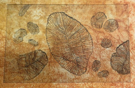
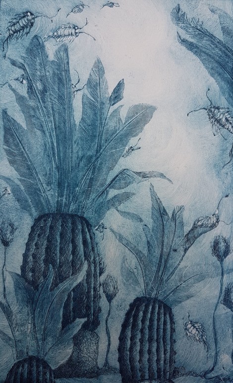
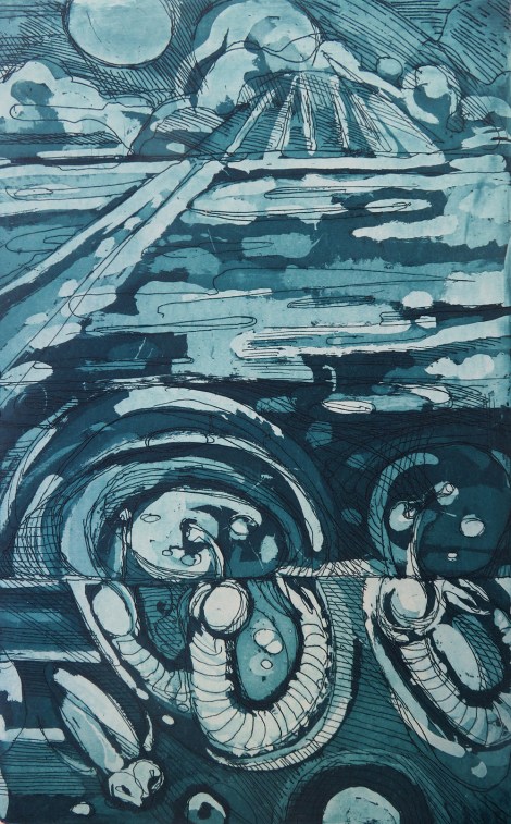
For our current exhibition, First Animals, we’ve taken this collaboration to a new level by commissioning original works from a total of 22 artists, all part of Oxford Printmakers Co-operative (OPC) – a group of over a hundred printmakers which has been running for more than 40 years.
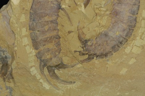
First Animals looks at the very earliest evidence of life on Earth, dating back half a billion years. Some of the fossils on display are shallow impressions in the rock – the only direct evidence we have that life existed at that time.
Amplectobelua symbrachiata – one of the incredible Cambrian fossils from the Chengjiang site in China
To kick-start the project we ran a series of workshops for OPC artists to meet the Museum researchers working on the exhibition, and to see the fossils first hand. There were also opportunities to draw directly from these unique fossils, many of which have never been displayed in the UK before.
Discussions between researchers and artists revealed fascinating similarities between these ancient fossils and the process of printmaking. Sally Levell, of Oxford Printmakers Co-operative, explains:
I was completely fascinated by the fossil collection in the Museum, especially the fine specimens from Chengjiang and Newfoundland. They are preserved as mere impressions in the rock, so they are, in essence, nature’s prints.
Each printmaker partnered with a researcher who could answer questions, provide extra info and help the artist decide which specimen or subject to depict in their final print. It’s clear from talking to the printmakers that this direct contact with the experts was invaluable and made the work really meaningful.
Xianguangia by Charlie Davies
We couldn’t have worked without the patient explanations and “show and tell” sessions with the three main researchers – Dr Jack Matthews, Dr Imran Rahman and Dr Duncan Murdock. They were just excellent and their dedication to their work was an inspiration to all of us printmakers.
Sally Levell
Over a period of around seven months, ideas blossomed and printing presses were put into action, with the printmakers exploring the forms, textures and evolution of the fascinating first animals. The final result is First Impressions, an enticing art trail of twenty-five prints dotted around the Museum, both within the First Animals exhibition gallery and nestled within the permanent displays.
Ottoia by Jackie Conway
Such a large group of artists brings a huge variety of techniques and styles, all under the umbrella of printmaking; from a bright, bold screen print in the style of Andy Warhol, to a delicate collagraph created from decayed cabbage leaves! To take part in the art trail yourself, simply grab a trail map when you’re next in the Museum.
Workshop printers inking up their plates
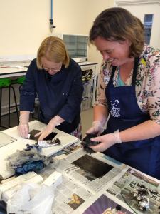
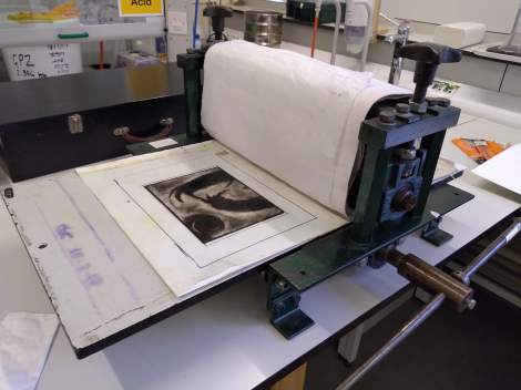
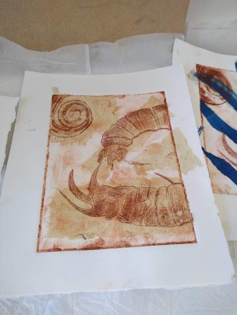
But our foray into fossils and printmaking didn’t stop there. OPC member Rahima Kenner ran a one-day workshop at the Museum where participants made their own intaglio prints inspired by the First Animals fossils. The group of eight people featured artists and scientists alike, all keen to capture the unique fossils through print techniques.
Designs were scratched onto acrylic plates and inked up, before a professional printing press created striking pieces to take home. Participants also explored techniques such as Chine-Collé, the addition of small pieces of paper to create texture and colour underneath the print.
It was a delight to be able to share with the group our enthusiasm for these discoveries in the medium of making the drypoint prints and to share their enjoyment of learning and using the new techniques. Some lovely work was produced in a single day.
Rahima Kenner
A plate about to go into the press
A finished print, using intaglio and chine-colle
The First Impressions project has been transformative for the Museum team and for the Oxford Printmakers Co-operative. Catriona Brodribb describes its impact on the printmakers :
It’s been a great opportunity to challenge one’s own artistic boundaries in terms of stretching the imagination, and for our members to throw themselves into something new, and enjoy responding to such ancient material in a contemporary way.
The First Animals and First Impressions exhibitions are open until 24 February 2020 and are free to visit.


")
")

")
")
")


")
")