First Impressions: exploring early life through printmaking

Dickinsonia by Claire Drinkwater

by Rachel Parle, public enagement manager

In each of our special exhibitions, we complement contemporary scientific research with contemporary art. In recent years this has included Elin Thomas’s crocheted petri dishes, Ian Kirkpatrick’s migration and genetics-themed installation, and who could forget the enormous E. coli sculpture by Luke Jerram?!

First Animals exhibition is on show until 24 February 2020

For our current exhibition, First Animals, we’ve taken this collaboration to a new level by commissioning original works from a total of 22 artists, all part of Oxford Printmakers Co-operative (OPC) – a group of over a hundred printmakers which has been running for more than 40 years.

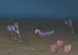
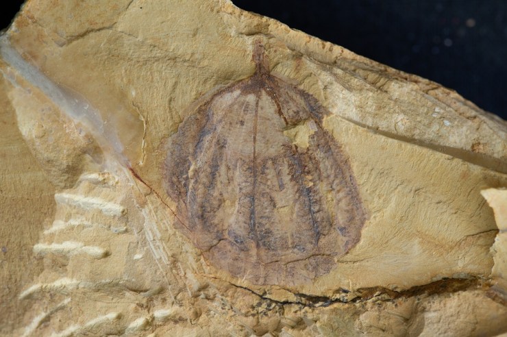
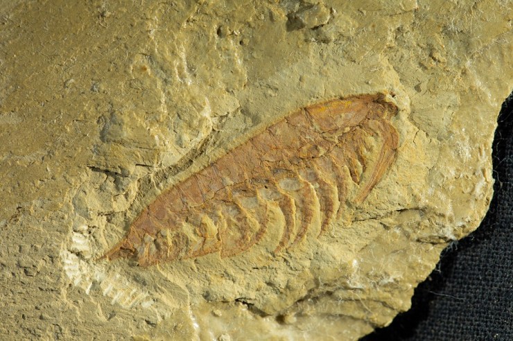
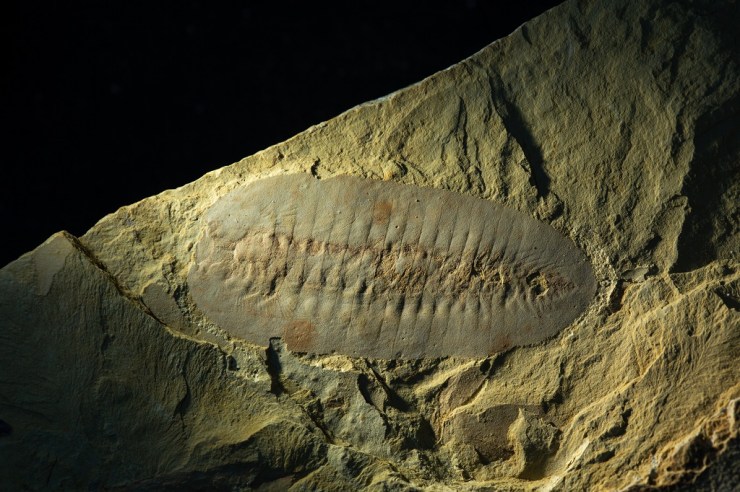
First Animals looks at the very earliest evidence of life on Earth, dating back half a billion years. Some of the fossils on display are shallow impressions in the rock – the only direct evidence we have that life existed at that time.

Amplectobelua symbrachiata – one of the incredible Cambrian fossils from the Chengjiang site in China

To kick-start the project we ran a series of workshops for OPC artists to meet the Museum researchers working on the exhibition, and to see the fossils first hand. There were also opportunities to draw directly from these unique fossils, many of which have never been displayed in the UK before.

Discussions between researchers and artists revealed fascinating similarities between these ancient fossils and the process of printmaking. Sally Levell, of Oxford Printmakers Co-operative, explains:

I was completely fascinated by the fossil collection in the Museum, especially the fine specimens from Chengjiang and Newfoundland. They are preserved as mere impressions in the rock, so they are, in essence, nature’s prints.

Each printmaker partnered with a researcher who could answer questions, provide extra info and help the artist decide which specimen or subject to depict in their final print. It’s clear from talking to the printmakers that this direct contact with the experts was invaluable and made the work really meaningful.

Xianguangia by Charlie Davies

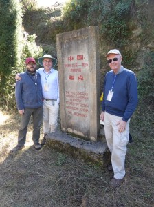
We couldn’t have worked without the patient explanations and “show and tell” sessions with the three main researchers – Dr Jack Matthews, Dr Imran Rahman and Dr Duncan Murdock. They were just excellent and their dedication to their work was an inspiration to all of us printmakers.

Sally Levell

Over a period of around seven months, ideas blossomed and printing presses were put into action, with the printmakers exploring the forms, textures and evolution of the fascinating first animals. The final result is First Impressions, an enticing art trail of twenty-five prints dotted around the Museum, both within the First Animals exhibition gallery and nestled within the permanent displays.

Ottoia by Jackie Conway

Such a large group of artists brings a huge variety of techniques and styles, all under the umbrella of printmaking; from a bright, bold screen print in the style of Andy Warhol, to a delicate collagraph created from decayed cabbage leaves! To take part in the art trail yourself, simply grab a trail map when you’re next in the Museum.

Workshop printers inking up their plates

But our foray into fossils and printmaking didn’t stop there. OPC member Rahima Kenner ran a one-day workshop at the Museum where participants made their own intaglio prints inspired by the First Animals fossils. The group of eight people featured artists and scientists alike, all keen to capture the unique fossils through print techniques.

Designs were scratched onto acrylic plates and inked up, before a professional printing press created striking pieces to take home. Participants also explored techniques such as Chine-Collé, the addition of small pieces of paper to create texture and colour underneath the print.

It was a delight to be able to share with the group our enthusiasm for these discoveries in the medium of making the drypoint prints and to share their enjoyment of learning and using the new techniques. Some lovely work was produced in a single day.

Rahima Kenner

A plate about to go into the press
A finished print, using intaglio and chine-colle

The First Impressions project has been transformative for the Museum team and for the Oxford Printmakers Co-operative. Catriona Brodribb describes its impact on the printmakers :

It’s been a great opportunity to challenge one’s own artistic boundaries in terms of stretching the imagination, and for our members to throw themselves into something new, and enjoy responding to such ancient material in a contemporary way.

The First Animals and First Impressions exhibitions are open until 24 February 2020 and are free to visit.

Abigail Harris - artwork showing reconstruction of Cambrian ocean animal life

Cambrian creation

Abigail Harris - artwork showing reconstruction of Cambrian ocean animal life

by Abigail Harris

Over the past few months our researchers have been working with University of Plymouth illustration student Abigail Harris, who has delved into the weird and wonderful world of some of the earliest animals. Here, Abigail tells us about the process that led to the creation of her Cambrian artwork, inspired by our First Animals exhibition.

I first visited the Museum in April this year when I was given the opportunity to collaborate with scientists as part of a module in my BA in at the University of Plymouth. Things kicked off with a short talk about the Ediacaran and Cambrian geological periods, when Earth’s first animal life started to appear.

I quickly narrowed my interest down to fossils from the Cambrian period which are more complex life forms, more similar to life today. A collection of small fossils from the Chengjiang fossil site in Yunnan province, China was the inspiration for some initial observational drawings.

Abigail Harris - sketches for artwork showing reconstruction of Cambrian ocean animal life
A sketchbook page showing initial sketches and observations of Onychodictyon
Final illustration of Cotyledion

After returning to Plymouth University, I began to develop these initial sketches and observations, continuing to research the Chengjiang material and learning more about the characteristics of some of the creatures preserved as fossils.

I wanted to create an under-the-sea ecology reconstruction showing a diversity of life forms, focusing on Onychodictyon, Cotyledion, Cricocosmia, Luolishania, and Paradiagoniella.

A five-step process was used for each reconstruction. Initially, I would sketch the fossil as I saw it, then I would research the characteristics and features of that animal, making a list of things to include in my drawing. A second drawing would then include all of these characteristics, not just what was initially visible in the fossil.

These rough sketches were then sent to the scientists for feedback, helping me to redraw and paint the illustrations with watercolour, before scanning and digitally editing each painting. Lastly, I created a background and added my illustrations.

Initial under under the sea ecology reconstruction.

Although the reconstructions were not completely finished by the time of my project deadline, I returned to the Museum in July and was given a tour of the First Animals exhibition by Deputy Head of Research Imran Rahman, as well as the opportunity to discuss how to improve my artworks for accuracy.

Another round of sketching and painting led to the final piece, shown at the start of this article, complete with an added digital background of the seafloor, and darkened to reflect the murky world of a Cambrian ocean, 50 metres below the surface.

Exceptional Chinese fossils come to Oxford in new partnership

by Imran Rahman, Deputy Head of Research

China is world-famous for its unique and exceptionally preserved fossils, which range from some of the oldest animals on Earth, to spectacular feathered dinosaurs. We are therefore very excited to announce that the Museum, along with other institutions from across Europe, is a partner in a major new venture with Yunnan University in China: the International Joint Laboratory for Palaeobiology and Palaeoenvironment.

Collaboration between this Museum and Yunnan University dates back to the 1990s, driven by the work of Professor Derek Siveter – a former Senior Research Fellow and current Honorary Research Associate at the Museum. He collaborated with Professor Hou Xianguang, director of the International Joint Laboratory for Palaeobiology and Palaeoenvironment, to study fossils from the internationally renowned Chengjiang biota, which was discovered by Hou Xianguang in 1984.

Museum researchers Duncan Murdock, Jack Matthews and Derek Siveter (l-r) visit the Precambrian-Cambrian Section

The Chengjiang fossil site is important and exciting because it preserves both the soft and hard parts of a range of early animals. This fossil record captures the rapid diversification of life about 520 million years old – in an event referred to as the Cambrian explosion. Derek Siveter was instrumental in a successful bid to have the Chengjiang biota designated a UNESCO World Heritage site in 2012, preserving it for future generations.

In December 2018, Museum researchers Duncan Murdock, Imran Rahman and Jack Matthews travelled with Derek to Kunming, China, for the first meeting of the International Joint Laboratory for Palaeobiology and Palaeoenvironment. The lucky researchers spent three days on field trips to the region’s most spectacular fossil sites, including Lufeng World Dinosaur Valley and the Chengjiang biota itself, followed by two full days of scientific talks and discussions.

The International Joint Laboratory is funded by the Ministry for Education of China and includes the University of Leicester, the Natural History Museum, London, the University of Munich, and the Bavarian State Collection of Zoology, along with Oxford University Museum of Natural History and Yunnan University.

The arthropod Haikoucaris ercaiensis. Sometimes referred to as a ‘short-great-appendage’ arthropod, Haikoucaris had a pair of prominent grasping appendages adjacent to the head (right-hand side of this image). Credit: Scott Billings
The arthropod Leanchoilia illecebrosa. Sometimes referred to as a ‘short-great-appendage’ arthropod, Leanchoilia illecebrosa had a pair of prominent grasping appendages adjacent to the head (right-hand side of this image). Credit: Scott Billings

A significant first outcome of this new partnership will be the loan of iconic Chengjiang fossil specimens from Kunming to Oxford for our First Animals exhibition which opens on 12 July and runs until February 2020. Most of these fossils have never been outside of China before, and some have never been seen by the public before. We invite you to visit First Animals to see these exceptional fossils first hand!

The arthropod Saperion glumaceum. Saperion had a flattened, segmented body and jointed appendages (not visible in this specimen). Credit: Scott Billings.

Top image: The comb jelly Galeactena hemispherica. Unlike modern comb jellies, which are soft-bodied animals, Galeactena and its relatives had hardened ‘spokes’ on the sides of the body (appearing as dark bands in this photograph). Credit: Scott Billings.

Animating the extinct

This sumptuous video features on our brand new Out of the Deep display and brings to life the two large marine reptile skeletons seen in the cases. The Museum exhibition team worked with Martin Lisec of Mighty Fossils, who specialise in palaeo reconstructions. Martin and his animators also created a longer video explaining how the long-necked plesiosaur became fossilised, as well as beautiful illustrations of life in the Jurassic seas. 
Martin explains the process of animating these long-extinct creatures:

The first step was to make 3D models of all the animals that would appear in the films or illustrations. After discussion with the Museum team, it was clear that we would need two plesiosaurs (one short-necked, known as a pliosaur, one long-necked), ammonites, belemnites and other Jurassic sea life. Now we were able to define the scale of detail, size and texture quality of the model.

In consultation with Dr. Hilary Ketchum, the palaeontologist on the project, we gathered important data, including a detailed description of the discovered skeletons, photographs, 3D scans, and a few sketches.

This slideshow requires JavaScript.

We created the first version of the model to determine proportions and a body shape. After several discussions with Hilary, some improvements were made and the ‘primal model’ of the long-necked plesiosaur was ready for the final touches – adding details, mapping, and textures. We could then move on to create the other 3D models.

This slideshow requires JavaScript.

The longer animation was the most time-consuming. We prepared the short storyboard, which was then partly changed during the works, but that is a common part of a creative job. For example, when it was agreed during the process that the video would contain description texts, it affected the speed and length of the whole animation – obviously, it has to be slower so that people are able to watch and read all important information properly.

A certain problem appeared when creating the short, looped animation. The first picture had to precisely follow the last one – quite a difficult goal to reach in case of underwater scenery. Hopefully no-one can spot the join!

This slideshow requires JavaScript.

At this moment we had a rough animation to be finalised. We had to make colour corrections, add effects and sound – everything had to fit perfectly. After the first version, there were a few more with slight adjustments of animation, cut and text corrections. The final version of both animations was ready and then rendered in different quality and resolution for use in the display and online.

The last part of the project was creating a large illustration, 12,000 x 3,000 pixels, which would be used as a background for a large display panel. Text, diagrams and a screen showing the animations would be placed on this background, making the composition a little tricky. We agreed that the base of the illustration would be just the background. The underwater scene and creatures were placed in separate layers so that it would be easy to adjust them – move them, change their size, position etc.

This slideshow requires JavaScript.

In the first phase, we had to set the colour scale to achieve the proper look of the warm and shallow sea, then we made rough sketches of the scene including seabed and positions of individual creatures. We had to make continuous adjustments as the display design developed.

Then we finished the seabed with vegetation, gryphaea shells and plankton floating in the water. The final touch was to use lighting to create an illusion of depth for the Jurassic creatures to explore.

*

More Out of the Deep videos are available on the Museum website.

Pieces of a plesiosaur

We’ve just opened a brand new, permanent display called Out of the Deep, featuring two beautifully preserved plesiosaur skeletons. Remarkably, both of these marine reptile fossils have skulls, which is more unusual than you might think. Dr Hilary Ketchum, collections manager in the Museum’s Earth Collections and curator of Out of the Deep, describes how the skull of the long-necked plesiosaur made it safely from a quarry to a museum display.

At the bottom of a clay pit in 2014, palaeontologists from the Oxford Clay Working Group discovered a 165-million-year-old fossil plesiosaur skeleton, and they knew they had found something special. Plesiosaur bones are fairly common in the quarry, but skeletons are rare. Skeletons with skulls are rarer still. Fantastically, at the end of their newly-found plesiosaur’s neck was a skull. Barely visible underneath the clay, only the tip of the snout and a few teeth were exposed.

Can you see the skull? Fossil hunting in the quarry takes time, patience and a good eye to distinguish between bones and clay. Image: Mark Wildman, Oxford Clay Working Group.

Plesiosaur skulls are usually made up of around 33 bones, not including the tiny bones from inside the eye sockets, called the sclerotic ring. The skull bones are among the smallest and most fragile in the entire skeleton. This means they are much less likely to be preserved, and less likely to be discovered, than the larger and more robust backbones and limb bones.

A plaster jacket was made around the skull while still in the quarry.
Image: Mark Wildman, Oxford Clay Working Group.

When the plesiosaur skeleton arrived in the Museum in 2015, the skull and some of the surrounding clay was encased in its protective plaster field jacket. As tempting as it was, instead of cracking open the jacket straight away, we decided on a more technological approach. Professor Roger Benson and Dr James Neenan took the specimen to the Royal Veterinary College to use their enormous CT scanner, normally used for scanning horses and other large animals, and took thousands of X-rays of the jacket. This allowed them to build up a 3D model of the fossil inside – our first tantalising glimpse of the whole skull!

The CT scan of the plaster jacket (left) revealed the location of the skull inside the jacket (middle). The jacket was then digitally removed (right) to reveal a 3D image of the skull.

Having the CT scan of the skull was like having a picture on a puzzle box
Juliet Hay, Earth Collections conservator and preparator

Although the CT scan was incredibly useful, we still had to proceed with the preparation with caution. It was possible that not all of the bones had not been detected by the scanner, especially the incredibly thin bones of the palate.

After opening the plaster jacket, Juliet began to carefully remove the clay from around the fossil bone.

Slowly and carefully, Juliet and I removed the soft clay from around the skull. The weight of clay pressing on top of the skull for millions of years had crushed it, breaking some of the bones into a lot of smaller pieces. In order to keep track of them we attached a number to each piece of bone and photographed it from several different angles before removing it from the jacket.

Each individual bone was mapped using a numbering system. The numbers were attached with the conservation adhesive Paraloid B72 in acetone, so that they could be easily removed later.
The plesiosaur’s pointed teeth being revealed.

When all the bones had finally been removed from the clay, we had over 250 pieces. Next came the challenge of the three-dimensional jigsaw!

With knowledge about plesiosaur skulls from my PhD, and some extra expert help from Roger Benson and Dr Mark Evans, Curator of Natural Science and Archaeology, New Walk Museum and Art Gallery, I was able to build up the skull, piece by piece, until it was nearly whole again.

After many months of painstaking work, the beautifully preserved skull of this long-necked plesiosaur can finally be seen in the Out of the Deep display.

Amazingly, the skull is even more complete and more beautifully preserved than we could tell from the CT scan. The sutures between the individual bones can be seen in exquisite detail, and even though I work with fossils every day, I still find it amazing that it is 165 million years old.

*

With special thanks to:

Oxford Clay Working Group: Mark Wildman, Carl Harrington, Shona Tranter, Cliff Nicklin, Heather Middleton, and Mark Graham, who uncovered and excavated the long-necked plesiosaur.

Forterra, for generously donating the plesiosaur skeleton to the Museum, after it was discovered in a Forterra quarry. 

Life’s big bang?

by Harriet Drage and Scott Billings

You may have heard of the Cambrian Explosion, an ‘event’, starting roughly 540 million years ago, when all the major animal groups suddenly appear in the fossil record, an apparent explosion of life and evolution.

But was there really an evolutionary explosion of all these animal groups, or is the lack of evidence from earlier periods due to some peculiarity of the fossilisation process? The debate has rumbled on for a number of years.

Now, a new study from our research team, the University of Oxford’s Department of Zoology, and the University of Lausanne, claims that the early Cambrian saw the origins and evolution of the largest and most important animal group on Earth – the euarthropods – in a paper which challenges two major pictures of animal evolution.

Euarthropoda contains the insects, crustaceans, spiders, trilobites, and a huge diversity of other forms alive and extinct. They comprise over 80 percent of all animal species on the planet and are key components of all of Earth’s ecosystems, making them the most important group since the dawn of animals over 500 million years ago.

Exceptionally preserved soft-bodied fossils of the Cambrian predator and stem-lineage euarthropod Anomalocaris canadensis from the Burgess Shale, Canada. Top left: Frontal appendage showing segmentation similar to modern-day euarthropods. Bottom right: Full body specimen showing one pair of frontal appendages (white arrows) and mouthparts consisting of plates with teeth (black arrow) on the head. Images: A. Daley.

A team based at the museum, and now at Lausanne, conducted the most comprehensive fossil analysis ever undertaken on early euarthropods, to try and establish whether these animals really did emerge in the early Cambrian period, or whether fossilisation just didn’t occur in any earlier periods.

In an article published today in the Proceedings of the National Academy of Sciences they show that, taken together, the total fossil record does show a gradual radiation of euarthropods during the early Cambrian, 540-500 million years ago, challenging other ideas that suggest either a rapid explosion of forms, or a much slower evolution that has not been preserved in the fossil record.

Each of the major types of fossil evidence has its limitation and they are incomplete in different ways, but when taken together they are mutually illuminating
Professor Allison Daley

Reconstruction of the Cambrian predator and stem-lineage euarthropod Anomalocaris canadensis, based on fossils from the Burgess Shale, Canada. Reconstruction by Natalia Patkiewicz.

By looking at a huge range of fossil material the researchers ruled out the possibility that Pre-Cambrian rocks older than around 541 million years would not have preserved early euarthropods. The only plausible explanation left is that the origins of this huge animal group didn’t evolve until about 540 million years ago, an estimate which also matches the most recent molecular dating.

The timing of the origin of Euarthropoda is very important as it affects how we view and interpret the evolution of the group and its effects on the planet. By working out which groups developed first we can trace the evolution of physical characteristics, such as limbs.

Exploring all the evidence like this allows us to make an informed estimate about the origins of key animal groups, leading to a better understanding of the evolution of early life on Earth.

Model of the Cambrian stem lineage euarthropod Peytoia, based on fossils from the Burgess Shale. Top left: Closeup of the mouth parts and frontal appendages. Bottom right: Overall view of the body. Model and image: E. Horn.