More than mimicry

figure_3
Hunting for Bates specimens. The labels with pink strips tell us they were part of Bates’ collection.

A small exhibition has popped up on the upper gallery of the Museum, showcasing natural historian Henry Walter Bates. He’s famous for his theory on mimicry, but, as exhibition curator Gina Allnatt explains, there’s a lot more to discover about Bates.

**

Charles Darwin and Alfred Russel Wallace are now widely recognised as the co-discoverers of the theory of evolution. They are both established figures in the natural history world, but it was a lesser known contemporary who gave them a missing piece of the puzzle.

A panel from the display is put together behind the scenes

Henry Walter Bates was born in Leicester on 8th February 1825. He was originally apprenticed to a hosiery manufacturer, but his passion for insects sent him on a completely different path in life. In 1844, Bates encountered Wallace in a library, and the two men found they shared a mutual love of nature. Bates introduced Wallace to the field of entomology (the study of insects) and it wasn’t long before the two were planning a joint expedition to the Amazon. They funded the expedition almost entirely through the sale of specimens they collected. Wallace returned to England after four years, but Bates remained in the Amazon for a further seven years. When he finally returned to England he had amassed a collection of over 14,000 insects. 8,000 of these were new to science.

LEPI4132b_heraldica_Bates_dorsal
Bates Type specimen from the Godman-Salvin collection

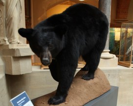
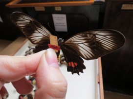
Bates is most famous for the work which bears his name: Batesian mimicry. Batesian mimicry is when a harmless species mimics the warning colours or behaviour of a harmful species.

The mimic  then benefits from the protection of the model. For example, the Hornet Moth (Sesia apiformis) is completely harmless, but looks like a wasp and benefits from the protection of the wasp’s warning colours. The moth is the mimic and the wasp is the model.

figure_2
Net cocoon (Urodidae) from the moth collections

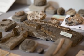
This Museum’s historical butterfly collection contains over 200 specimens collected by Bates, and thousands of other insects collected by him in the overall Entomology collections.

The specimens came to the collections via different routes. Some were purchased directly from Bates by Professor J.O. Westwood, the Museum’s first Hope Professor of Zoology. Bates material also arrived through acquired collections, such as those purchased from natural history specimen dealer Samuel Stevens. Other specimens of interest in the Lepidoptera collections purchased from Bates include an unusual net cocoon from a rare moth in the family Urodidae. The Urodidae are an unusual form of moth that build cocoons resembled a mesh bag with an opening at the bottom. One theory for this unusual structure is  to allow rainwater to flush through it easily, without drowning the pupa inside. In the Amazon rainforest, which is prone to heavy rainfall and flooding, this is a huge advantage.

The exhibition, which can be seen until 26th February 2016, will reveal much more about Bates and his contribution to modern-day science.

Gina Allnatt, Curatorial assistant (Lepidoptera)

Talkin’ ’bout taxidermy

IMG_1691 (1)

Here at the Museum we have many bird, mammal, fish and reptile taxidermy specimens on display. There are a fair few that you can touch too, on our Sensing Evolution tables in particular. Outside museums, taxidermy has seen a wider surge in popularity in recent years, featuring in artistic practice and being offered as a practical skill through taxidermy courses. It has also been the subject of ethical questions and debates.

In the 19th century, taxidermy was seen as one of the ways of bringing strange creatures from the around world to museum visitors. They were collected at a time when the natural world was thought to be an infinite resource. Many early biologists would combine scientific expeditions with collecting trips, gathering material to be studied, described and displayed in public museums.

IMG_1639 (1)
Unwrapping the American Black Bear

Today, it’s no longer possible to simply order an animal from a collector and expect to receive a crate containing a specimen prepared for display. There is a raft of national and international legislation designed to ensure that biological material is ethically, legally and safely collected and transported. It is also concerned with the impact on source communities, species conservation or ecosystems, and imposes checks on the transmission of disease or pests.

When Sensing Evolution opened here in May 2015 the museum obtained two larger pieces of taxidermy to greet visitors as they enter the building: a Red Deer purchased from a farm in Belgium, and an American Black Bear, which is a donation from the New Jersey Fish and Wildlife department in the USA.

Commissioning taxidermy is not straightforward and it’s important to know which permits are needed and whether you can legally qualify to receive them. There are many laws surrounding the import and use of animal parts that you need to be familiar with.

The Museum believes in the importance of wildlife conservation and we wanted to be sure that our taxidermy specimens are ethically obtained. Neither the Red Deer nor the American Black Bear were killed for the purpose of museum taxidermy, and both were sourced following a lot of research and communication with professional taxidermists and zoological societies.

IMG_1674 (1)
Team Bear and the bear itself, now installed in the court

The Red Deer skin was discarded from a farm where the specimen was raised for meat; the Black Bear was euthanized by the New Jersey Fish and Wildlife department as part of its standard control measures, although in this case rather than incinerating the carcass, the skin was removed, processed and donated to the Museum. We waited for many months for a suitable specimen to become available in this way.

The result is two touchable pelts which we hope visitors will find fascinating and beautiful, and which will contribute to our understanding of these incredible animals.

For more advice or information about taxidermy contact your local museum or the Guild of Taxidermists.

It’s offishal!

Tuna 1

This skeleton has been on Bethany Palumbo’s conservation hit list for some time. As the Conservator for our Life Collections, she keeps a close eye on specimens that need a bit of TLC, or indeed a full make-over. Bethany and team have now been given the go-ahead to dedicate their attention to the Atlantic Bluefin tuna (Thunnus thynnus), located in the Museum court. The project will kick off on November 9th and should take around 4 weeks to complete.

This specimen has a long and interesting history. It’s been on display in the Museum since it opened in 1860, but records show that it was previously displayed in the Anatomy Museum at Christ Church College, Oxford. The fish was collected by Dr. Henry Acland, a leading figure in the establishment of the Museum. He obtained it through the Consul of Maderia in 1847 and its remarkable story was recorded in a historical review of the Museum Zoological collections (1976).

‘…the fish had been packed in salt and placed in an eight foot-long box addressed to ‘Dr Acland, Oxford’. During the voyage, the crew and passengers had become convinced that the box contained the corpse of a patient which in their superstitious minds was sufficient to account for the storm in the Bay of Biscay. The crew and passengers could not be convinced otherwise and Acland submitted to the opening of the ‘coffin’ by the ship’s carpenter, who unscrewed the lid before the assembled company, to reveal the tunny for all to see. It was eventually delivered to the Museum at Christ Church in perfect condition, the skeleton being later carefully articulated by Charles Robertson’.

Conservation intern Abby assesses the condition of the Bluefin Tuna in order to develop a plan for treatment.

Now classified as an endangered species, this Bluefin Tuna also has significant scientific, as well as historical importance. Our conservators will take this into account, ensuring that any treatment is carried out sensitively. To allow Museum visitors to watch conservation in action, the work will be carried out right in the Museum court. Bethany is looking forward to lots of conversations with the public about all things fishy!

Rachel Parle, Interpretation and Education Officer

Layer upon layer

IMG_6299

Handwritten in Stone
9 October 2015 – 31 January 2016

Today sees the opening of our new special exhibition – Handwritten in Stone – celebrating the life and work of William Smith on the bicentenary of his publication of the first geological map of England and Wales.

Dubbed ‘The Map that Changed the World’ in Simon Winchester’s book of that title, the beautifully hand-coloured map revealed a three-dimensional arrangement of rock layers, or strata, along with a fourth dimension – time.

This work earned Smith the moniker ‘the father of geology’, an accomplishment all the more impressive given that Smith achieved it single-handedly and with very little formal education.

This 1799 map of Bath, on display in the exhibition, is the oldest geological map in the world

The Museum holds the largest archive of Smith material in the world. Alongside the famous 1815 map, shown at the top of the article, are personal papers, drawings, publications, maps and geological sections, most of which are being displayed for the first time. With these we have some fossil material from the collections: Smith realised that particular combinations of fossils were unique to different rock formations and could be used to date the strata.

Design work on the 'drawing board', created by Claire Venables at Giraffe Corner
Design work on the ‘drawing board’, created by Claire Venables at Giraffe Corner. Photo: Claire Venables

Handwritten in Stone, supported by the Heritage Lottery Fund, is the first show in our new special exhibition gallery on the upper east side of the building. Working out how to use the space was a learning process, and the final, elegant design is the work of local consultancy Giraffe Corner.

To bring the final exhibition together we collaborated with writer Rebecca Mileham, an installation team from the Ashmolean Museum, and more than 50 volunteers. Look out for more about the volunteers’ contributions on this blog soon.

Applying the graphics in the gallery
Applying the graphics in the gallery. Photo: Claire Venables

The 1815 map itself takes centre-stage in the exhibition, flanked on one side by the history of Smith’s work building up to its publication, and on the other by the legacy of his techniques, which are still used today.

To discover the full story of William ‘Strata’ Smith head over to the upper east side of the Museum before 31 January.

And don’t forget to look out for our What’s On programme which includes lots of William Smith and geology-focused events, including a public talk by author Simon Winchester on 13 October.

Scott Billings – Public engagement officer

Presenting… the Gault Clay

IMG_5120

Our changing Presenting… display offers a look at highlights, themes and topical material drawn from the Museum’s collections. The current display presents a rich selection of fossils taken from a formation known as the Gault Clay. This will be on show until 21 September (Madagascar is up next), so it’s almost your last chance to see it.

The Gault Clay is known for its exceptional diversity of fossils. Deposited across northern Europe in the Lower Cretaceous, 100-112 million years ago, its abundance of ammonite fossils shows that it was laid down in a marine environment, at a time when sea levels were rising rapidly.

The Gault Clay in the Cliffs at Folkestone

Best seen at Folkestone in Kent, the Gault is sandwiched between the Lower Greensand and Chalk formations. The Folkestone Warren landslip has provided ample opportunities for fossil collectors for centuries. The land is still on the move today, but 100 years ago, on 19 December 1915, a major slip occurred which shut down the local railway for five years.

IMG_5122
Ammonite (Hoplites dentatus)

William Smith (1769-1839), who published the first geological map of England and Wales 200 years ago this year, called the Gault ‘blue marl’. He used the fossils found in it to map the Gault across the South of England.

Along with ammonites, it is possible to find snails, clams, shark’s teeth, fish, crabs, and lobsters. Even dinosaur fossils have been found, including the ankylosaur Anoplosaurus.

Some of the Gault collections in the Museum date back to the 1800s; others were collected as recently as 2015.

The Presenting... display, near the entrance to the Museum
The Presenting… display, near the entrance to the Museum

Bringing dinosaurs to life

Iguanodon bernissartensis

By Rachel Parle, Education and Interpretation Officer

Dinosaurs were living, breathing, moving animals, but that’s sometimes hard to visualise when standing in front of a skeleton. We may not be able to reincarnate dinosaurs in the style of Jurassic World, but an excellent illustration of the animals in their environment can go a long way to bringing them back to life.

Triceratops horridus

When Earth Collections Manager Hilary Ketchum and I set out to update the labels for our free-standing dinosaur skeletons, we wanted to present current science alongside scientifically accurate illustrations. They should be beautiful and show the dinosaurs as dynamic animals. We found just the person for the job. Julius Csotonyi is a paleoartist, wildlife artist and scientific illustrator who specialises in life-like restorations of prehistoric animals and habitats. He understood exactly what we wanted and set to work researching featured specimens.

Pachycephalosaurus wyomingensis

After several rounds of checks and suggestions from scientists in the Museum and the University, the illustrations are all complete and the new labels are on display in the Museum. So I asked Julius a few questions about his work and how he felt having completed the project.

**

Julius working on a dinosaur reconstruction.
Julius working on a dinosaur reconstruction. Credit: Alexandra Lefort

How do you ensure your representations of dinosaurs are accurate?

Ultimately, all of my reference material comes from palaeontologists’ research. For all reconstructions, I rely heavily on published scientific literature. For reconstructions of newly discovered taxa, of which I am commissioned to do quite a few for press releases and scientific papers, I also have discussions with the palaeontologists who have made the discoveries, since the material is not yet published. This latter process is some of the most exciting, because I am able to participate in the process of scientific discovery, keeping a foot in both camps of science and art.

Struthiomimus sedens

How important do you think the dinosaur’s environment is in the representation?

The environmental context provides the opportunity to tell a more detailed story of the animal’s role in the biological community, its position in the food web, or interesting aspects of its behaviour. Depicting the animal’s environment provides me the opportunity to employ creative and interesting lighting conditions and composition to generate an image that is as aesthetically appealing as possible – this is art, after all, and I feel it’s important to make it as beautiful as I can.

How did you become a paleoartist and what do you enjoy about it?

The new Iguanodon on display at the dinosaur's feet
The new Iguanodon label on display at the dinosaur’s feet

I absolutely love my job. It’s wonderful to play a part in piecing together and visualizing worlds that are millions of times older than I am. It was during the completion of my PhD in the microbiology of extreme environments that my work in scientific illustration and paleoart really took off, when I was first contacted to help illustrate a book about dinosaurs by author Dougal Dixon. Ultimately I realized that scientific illustration provided me with a more consistent enjoyment, so I made scientific artwork my full time work as soon as I completed my degree. I know that I am in the right field of work because even when I am juggling projects under the extreme pressure of impending deadlines, I still find great enjoyment in the act of painting.

This up-to-the-minute T. rex has feathers!


How do you feel about your work being on permanent display here?

I feel greatly honoured to have my illustrations incorporated into a permanent display in this renowned and respected institution. It is my hope that my work will help in a small way to interest the public in the intriguing field of palaeontology, and this excites me, for I feel strongly about contributing to scientific outreach. Many thanks to the museum team for allowing me to participate!

**

Next time you call by the Museum, stand in front of a dinosaur, have a good look at its new label and see if it comes to life before your eyes.

Rachel Parle, Interpretation and Education Officer

All images are copyright Oxford University Museum of Natural History and Julius Csotonyi.