Museums are a place for many things: inspiration, learning, conservation… the list goes on. But we believe that they should also be a forum for debate and discussion. One of our aims, as part of our Contemporary Science and Society exhibition and event series, is to bring controversial or challenging ideas to our visitors and to encourage a lively, informed and balanced debate.
Thomas Henry Huxley
Controversy is nothing new to this institution; there is a history of debate going right back to 30 June 1860, the year the Museum was founded. The Great Debate is believed to have been the first ever public debate on Charles Darwin’s theory of evolution – certainly the thorny issue of the time.
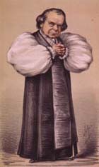
The debate is now notorious for the clash of ideologies between Samuel Wilberforce, the Bishop of Oxford, and Thomas Henry Huxley, a young biologist known as ‘Darwin’s bulldog’.
Samuel Wilberforce
Reports from the time are a little sketchy, but tempers are believed to have flared and insults were traded, climaxing with the shocking moment where Wilberforce compared Huxley’s grandparents to an ape. This was obviously outrageous to the delicate Victorian temperament, and people were believed to have fainted with shock!
This short video reveals how we think the debate may have gone (with a little artistic license thrown in):
The anniversary of the Great Debate falls next week, and this year it’s an extra special one. We’re reigniting the tradition of a good lively discussion with The Great Debate: Smart Drugs – Is It Cheating? On Thursday 29 June, Claire Fox of the Institute of Ideas and BBC Radio 4 chairs a multidisciplinary panel as in a debate about the ethics, fairness, and effects of so-called smart drugs and their impact on society.
Smart drugs is a name given to prescription drugs, typically used to treat disorders such as narcolepsy and attention deficit hyperactivity disorder (ADHD), which are now also commonly being used to improve cognitive ability and concentration. Some studies suggest that these drugs are now widely being used by university students, in a climate of increased academic and financial pressure.
Many students are said to see these drugs as a normal aid to study, but some experts have serious concerns about increasing levels of self-prescription and the long-term safety of their use, as well as the impact on competitiveness. Increased use in other areas of society may also have implications.
The panel for this debate includes world experts in the fields of neuroethics, evolutionary psychology, and philosophy, each representing different sides of this challenging subject.
Tickets are free, but you need to book your place. We can promise controversial opinions, expert insights and an eye-opening evening, but unlike Wilberforce and Huxley, we can’t necessarily guarantee that the panelists will be flinging insults about each other’s grandparents.
The latest display in our changing Presenting… case showcases a wonderful array of dung beetles. Darren Mann, head of our Life Collections, tells us why they are so important.
Worshipped during ancient Egyptian times, dung beetles have a long history of human appreciation. Jean-Henri Fabre (1823-1915), one of the first to popularise insects in his writings, began his Souvenirs entomologiques series with the Sacred Scarab, and even Charles Darwin appreciated the weaponry adorning many dung beetles.
Dung beetles can be divided into three main groups based on their nesting behaviour. The rollers, often seen on television wildlife documentaries, make a ball out of dung and roll it some distance before burying it. The tunnellers dig directly below the dung pile and bury as much as needed for nest construction. Finally, the dwellers nest within the dung pile.
The South American Phanaeinae is one of most colourful groups of beetles. They are often referred to as Rainbow Scarabs due to their bright metallic bodies. We don’t fully understand why these beetles are quite so colourful.
Dung beetles are one of the more popular groups of insects used in ecological and evolutionary research today. They can help us to understand questions about how biodiversity loss impacts on ecosystems, or act as model organisms in the field of evolutionary development.
Unlike the much-publicised importance of bees and their pollination services, dung beetles are relatively unknown, despite their huge ecological and economic value. Their feeding and nesting behaviours provide many useful ecosystem services such as dung removal, pest fly control, parasite suppression, nutrient cycling, plant growth enhancement, improvement of soil structure, secondary seed dispersal, and a possible reduction of greenhouse gas emissions.
Through these activities, one study calculated that dung beetles are worth around £367 million a year to the UK cattle industry alone.
The largest dung beetles belong to the genus Heliocopris, which can reach up to 69 mm (pictured is Heliocopris dominus). These large beetles specialise on elephant and rhino dung. From around 2 mm in length is the oriental genus Panelus. These small beetles probably feed on the ‘dung’ of other insects and fungi.
Ancient Egyptians believed that the dung beetle kept the Sun moving across the sky like a giant ball of dung, linking the insect to the god of the rising sun Khepri. Some historians believe that it was through observing dung beetle behaviour and biology that Egyptians developed ideas about life after death.
The two most widely depicted species in Egyptian art are Kheper aegyptiorum and Scarabaeus sacer. Nowadays, only Scarabaeus occurs in this region of Africa; Kheper is now a more southern species, possibly indicating climatic changes since Ancient Egyptian civilization.
Kheper aegyptiorum on display in the museum’s Presenting… case
The UK has about 60 species of dung beetle and most of these belong to the ‘lesser dung beetle’ subfamily Aphodiinae. The largest of our dung beetles are the Dor Beetles which can reach 28 mm. Our smallest, Plagionus arenarius, is a meagre 2.5 mm. Sadly, over 50 per cent of our dung beetles are in decline due to agricultural intensification, pesticides and habitat loss.
Each year the Museum works with members of the community on a wide variety of projects using our collections to enthuse and engage people in natural history. These projects often result in some amazing outcomes but until now we have been unable to find the right space to celebrate this work in the Museum. So this month we are very happy to unveil our new Community Case, dedicated to doing just that.
Stories from Stone, Body and Bone in the new community case
Our opening display focuses on the Children in Need-funded Story Makers programme. In partnership with Fusion Arts, this initiative helps Oxford primary school pupils to develop their communication skills by taking inspiration from museum collections. And this year they teamed up with us to create Stories from Stone, Body and Bone.
Pupils from New Marston, Wood Farm, and Rose Hill Primary schools worked with Story Makers founder and arts psychotherapist Helen Edwards in two visits to the Museum, stimulating and developing imaginative ideas, stories and artwork.
During these visits the Story Makers met with our education officer Chris Jarvis and together they looked at rocks and minerals, tectonic plate formation, and the evolution of skeletons and animal posture. They explored the collections creatively through sensory observation, using the hands, body and senses to develop self-awareness and self-confidence.
Getting creative with chalks and textiles
We work with the children as artists and we carefully designed a series of sessions that enabled them to have direct sensory engagement with objects in the museum. We then used art processes to portray their experiences and feelings about their interactions. Helen Edwards, Integrative Arts Psychotherapist
Back at school, the pupils used visual art, drama, movement and modelling to communicate feelings and ideas that emerged from these museum encounters, sharing thoughts with the group in a playful and trusting atmosphere.
Group sessions back at school involving movement, drama and art
Detail from one of the Stone Age caves
Each Story Maker then created a Stone Age character – someone who might dream up and pass on stories full of meaning and myth. They imagined places in which their Stone Age characters might live, thinking about what they might see looking out from these spaces, through the cracks, crevices and windows in their caves.
From these ideas emerged beautiful, bright, and colourful models of these fictional abodes, as well as stories and poetry about their characters.
Story Makers built the children’s capacity to think reflectively, enriching their speech and language, and helped them to develop their writing skills as the stories were compiled into Story Makers books.
Stone Age houses and landscapes as part of the Stories from Stone, Body and Bone project
Everyone should get to do this, it is like a dream come true Story Maker, from the Stories from Stone, Body and Bone project
Stories from Stone, Body and Bone is on display until Sunday 21 May in our new Community Case. The next display, installed on 22 May, will feature artwork by our community of artists who use the collections as inspiration for their work.
Can your brain rewire itself? How is the brain built and what can go wrong? And should the secondary school day start later to compensate for teenage sleep patterns?
Here we present a selection of videos from the exhibition. The full set is available on our YouTube channel now. And if you’re not able to visit the exhibition itself, we’ve built a special Brain Diaries website which contains all that neuroscience goodness.
Is brain-building a tricky business? Professor Zoltán Molnár of the University of Oxford specialises in the development of the brain. In this video he talks about the complex processes at play during the brain’s early development, including how things can sometimes go wrong.
Can my brain rewire itself? Associate Professor Holly Bridge works in the Nuffield Department of Clinical Neurosciences in the John Radcliffe Hospital in Oxford. Her research focuses on using MRI scans of the human brain to understand the organisation of the visual system in people with normal vision and in those with abnormal visual function. Here she talks about how the brain can rewire itself to compensate for damage to certain sensory areas.
School’s out – should the school day start later? Dr Christopher-James Harvey works at the Sleep and Circadian Neuroscience Institute at the University of Oxford. As part of the Teensleep research project, he is investigating how changes in the natural rhythm of sleep in adolescents, and the effects of sleep education, might impact on academic, health and sleep outcomes. Here he talks about initiatives to trial a later starting time for the secondary school day.
To read more about Brain Diaries and see the full programme of public events see braindiaries.org.
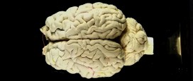
Our next exhibition – Brain Diaries: Modern Neuroscience in Action – opens on 10 March and in preparation we have indulged in a little bit of brain-washing… This article contains an image of a preserved human brain.
One of the first displays visitors will encounter is a ‘wall’ of 23 fluid-preserved mammal brains – from a Short-nosed Bandicoot to cow. The style of jar, with its black bitumen and paint backing, tells us that these were once used for display so it is exciting to put them in the public galleries again. Museum conservator, Jacqueline Chapman-Gray, runs us through the meticulous process she undertook to ensure these brains will look their best for their return to the limelight.
Cow brain before conservation treatmentA number of the brains had become dehydrated over time as the level of fluid – alcohol – had dropped. These needed to go through a rehydration programme to ensure their long-term preservation. This is more complex than simply adding more fluid to the jar. Instead the alcohol level needs to be increased gradually to avoid damaging the tissues.
Brains soaking in alcoholOthers had started to detach from their glass mounts, or anatomy labels that marked each of the different areas or sections of the brain had come loose. These were carefully remounted using specialist conservation-grade materials and a steady hand! Three brains had become completely detached and were repaired using a polyester monofilament thread, otherwise known as fishing line.
Repairing a human brain with a beading needle Labels found detached at the bottom of the jarFor the smallest of the brains a normal sewing needle was enough to pass through the tissues but for the larger two either a flexible 10cm beading needle or large 25cm mattress needle was needed. The original threading points were reused wherever possible though in one case this proved to be too difficult, as the tissue was soft and susceptible to breaking. With precision and patience I was able to gently stitch them back into place on the backing plate so they look as good as new.
All of the jars were given a thorough clean to ensure that seals were tight fitting and that the contents were shown off to their best. They were then filled with fluid to 4/5ths from the rim and the brains gently placed back inside.
Lids were sealed with clear silicone and each jar was topped up with a syringe through a small hole in the lid that is there for this very purpose – once full, this hole is also sealed.
Lastly, after the seals had dried, for the final finishing flourish black paint was reapplied to the backs and tops of the jars to provide a contrasting backdrop.
Ta-dah… the cow brain after conservation treatmentBrain Diaries opens on Friday 10 March and runs until Monday 1 January 2018. Take a look at the website to find out more about the exhibition and accompanying programme of events at braindiaries.org
The museum holds the only remaining soft tissue of the extinct dodo known anywhere in the world. The partially dissected skin of the head and scales from the feet of a single dodo represent one of natural history’s most iconic specimens. In fact, it is so tied to the museum’s identity and history that we use the dodo as our logo and it is even incorporated in the name of this blog.
Although the dodo head had been at Oxford University since the formation of the original Ashmolean Museum in the 17th century, it wasn’t really until the 19th century that the specimen really became celebrated.
Around this time, publications confirmed the extinction of the dodo from the island of Mauritius, where it was endemic. To capitalise on the rising interest in the animal, Ashmolean Museum Keeper John Duncan commissioned a number of casts of the Oxford Dodo head to give to, and exchange with, other museums.
One of the earliest of these casts was presented to the British Museum in 1828; later casts are recorded as being sent or exchanged with leading scientists of the time, as well as with Leiden Museum and the Royal College of Surgeons.
From these original and later casts further casts and models were presumably made, and eventually, dodo specimens spread to virtually every major natural history museum in the world. Today, many museums display casts of this head, all stemming from the single specimen held here in Oxford.
One of the many casts in the Oxford University Museum of Natural History, this one has been painted to match the original specimen
The Museum contains a number of models and casts of the head too; some are made from plaster and resin, some are painted to resemble the original specimen. The head of the dodo was actually dissected in 1847, by Henry Acland. He removed the skin from one side of the face so the early casts are a record of how the specimen would have looked originally.
In preparation for the Presenting display in the Museum I contacted natural history museums through the Natural Sciences Collections Association asking people to share information and photos about their casts and models of the dodo head. I wanted to try and construct a picture of how the dodo head was disseminated, as well as capture the diversity of quality and colours of representations of the original specimen. Here’s how far some of the dodos have flown:
Cast of the head labelled as coming from Cartwright Hall. Curator Gerry McGowan suspects this may have come via the Bradford Philosophical Society collections. The first curator of the society, Louis Compton Miall was friends with Thomas Henry Huxley and through him had contacts with many other geologists who may have gifted or exchanged this cast.
Bristol Museum & Art Gallery Bristol received a cast of the head directly from Oxford from Philip Duncan in 1834, keeper of the Ashmolean Museum between 1826 and 1855. Unfortunately, the head was likely destroyed in bombings of Bristol in 1940.
Canterbury Museum, New Zealand
Received a cast of a head from Professor Rolleston on 21 July 1871 in exchange for two kiwi skeletons which are still in the museum collections today.
Cast of head and foot presented to the museum by E.Ray Lankester in 1891/1892 just after leaving UCL and being appointed the Linacre Professor of Comparative Anatomy at Oxford.
The Great North Museum Hancock’s cast was presented by George Townsend Fox. This specimen had been presented to the Natural History Society of Newcastle in 1841 by Fox and had originally been presented by Philip Duncan.
Cast of the head that had quite a circuitous route to the Horniman Museum. The Horniman received the cast from the geology department of Queen Mary’s University of London in 1964 which received the cast from the Saffron Walden Museum in 1962.
Manchester Museum Cast of a head, presumed to have been presented by William Boyd Dawkins. The cast is currently on display in the Living Worlds gallery in Manchester Museum.
The National Geological Repository British Geological Survey
The Natural History Museum’s collections contain three casts of the Oxford Dodo head – two in the ornithology collections (pictured) and one in the palaeontology collections. The unpainted cast in the ornithology collections has the name ‘Johnson’ inscribed into the base.
Colleague Adam Smith got in touch with some interesting specimens from Wollaton Hall. The first one looks like another cast in this series but the second cast is unlike any of the others gathered here. The cast shows an open eye, detail on the beak as well as a more defined hook to the end of the bill.
Unfortunately, there’s not much information about the origins of these two casts so it’s probable that the ‘open eye’ cast may be a cast of a model reconstruction or an in progress sculpt. There’s an extremely slight chance it’s a cast of an otherwise unknown dodo head… If you recognise this dodo head do get in touch so we can solve this mystery for colleagues in Nottingham (it’s not the model dodo that we have on display here!).
Cast of a head at Warwickshire Museum with damage to the beak. Donated to the museum by clergyman and naturalist Reverand Andrew Bloxham in the 19th century. As the museum is currently moving stores, further information about when this cast was acquired is inaccessible.
**
If you work at a museum and have a dodo head cast to share, please do get in touch and we’ll update this blog accordingly.
Last updated: 10/11/17
‘Presenting… The Flight of the Dodo’ was on display at the Museum of Natural History from the 25 January to 22 March 2017.
Acknowledgements
With many thanks to colleagues across the sector who helped with information and images about dodo specimens: Adam Smith, Alice Adams, Jack Ashby, Carol Davies, Bonnie Griffin, Dan Gordon, Yvette Harvey, Mike Howe, Emma-Lousie Nicholls, Laura McCoy, Gerry McGowan, Nigel Monaghan, Henry McGhie, Pat Morris, Paul Scofield, Paul Shepherd, and Paul Sweet.


 The panel for this debate includes world experts in the fields of neuroethics, evolutionary psychology, and philosophy, each representing different sides of this challenging subject.