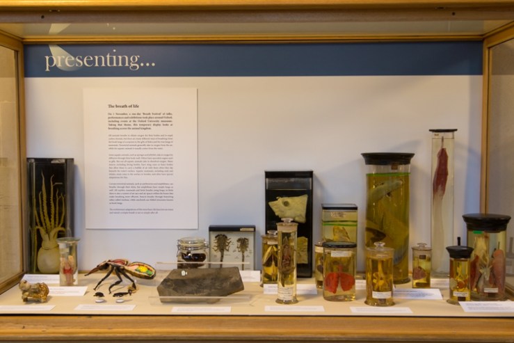
Tomorrow afternoon the Museum will host talks, tours and a dance performance as part of the Breath Festival, a unique series of events coordinated by the Oxford University Hospitals Artlink programme. To coincide with the festival we have put together a special display in our changing Presenting… case, all about breath and breathing across the animal kingdom.
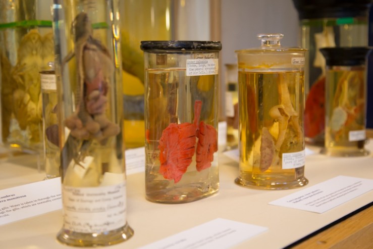
There’s something of the Halloween macabre about the display too, with its pink-coloured lungs and eviscerated bodies suspended in spirit. Here’s a taster of the display, but to see the full selection head down to the Museum either for the Breath Festival tomorrow, Saturday 1 November, or at any time during the rest of the month.
Lungs of a lizard, goose and duck
The breath of life All animals breathe to obtain oxygen for their bodies and to expel carbon dioxide, but there are many different ways of breathing: from the book lungs of scorpions to the gills of fishes and the true lungs of mammals. Terrestrial animals generally take in oxygen from the air, while for aquatic animals it usually comes from the water.
Crocodile and alligator lungs
Some aquatic animals, such as sponges and jellyfish, take in oxygen by diffusion through their body wall. Others have specialist organs such as gills. But not all aquatic animals take in dissolved oxygen. Many insects, including diving beetles, have wing cases or hairy bodies that allow them to carry a bubble of air with them when they dip beneath the water’s surface. Aquatic mammals, including seals and whales, must come to the surface to breathe, and often have special adaptations for this.
Certain terrestrial animals, such as earthworms and amphibians, can breathe through their skins, but amphibians have simple lungs as well. All reptiles, mammals and birds breathe using lungs; in birds there is also a system of air sacs and air spaces within the bones that make breathing more efficient. Insects breathe through branching tubes called tracheae, while arachnids use folded structures known as book lungs.
The evolutionary adaptations of this most basic life function are many and varied: a simple breath is not so simple after all.
According to the International Union for Conservation of Nature (IUCN) Lemurs are the most endangered mammals in the world. This diverse group of primates includes more than 100 species, but can only be found on Madagascar and the neighbouring Comoro Islands. On display here in the Museum we have a variety of lemurs, including the skeletons of extinct giant lemurs – some of which were as large as an adult human,
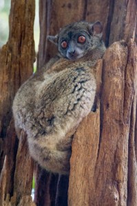
Sportive Lemur
With many species already extinct, the Lemur is in real trouble. This week a World Lemur Festival is being held in Antananarivo, the capital of Madagascar. To spread the word, the Lemur Conservation Foundation have put together a film to celebrate the festival and to highlight how close we are to losing the lemur.
Here in the UK, East Oxford residents and Museum visitors Tom Nicholson-Lailey and Janet McCrae approached us about the Lemur Festival. Janet and her partner Michael set up the McCrae Conservation and Education Fund in 2006 to support local conservation work in Madagascar. In collaboration with the late primatologist Dr Alison Jolly and Durrell Wlldlife, they’ve produced a series of posters, which have been distributed to 200 primary schools, featuring ecosystems under threat. Janet says she is
Mad about helping local children understand the unique habitat and their role in preserving it.
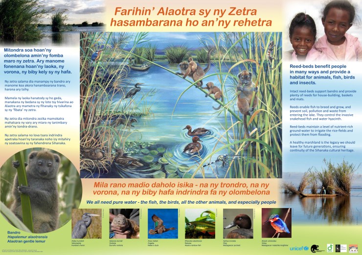
One of the posters designed by Janet Robinson for the McCrae Conservation and Education Fund
Tom has made 3 visits to Madagascar for wildlife-watching holidays, and has contributed the fantastic photographs you can see here. He says
“I hope that by travelling to Madagascar and visiting national parks and reserves, we are supporting local conservation work in Madagascar, and helping to ensure that conservation has a high political profile.”
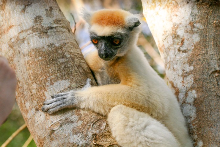
Golden Crowned Sifaka – one of the amazing photos by Tom Nicholson-Lailey
Madagascar, the fourth largest island in the world, is one of nature’s great treasure-houses, described as a ‘biodiversity hotspot’ for its number and variety of species. Some 90% of all the island’s animal and plant species are ‘endemic’ – found nowhere else in the world. The ‘flagship’ species that best represent Madagascar’s extraordinary biodiversity are the lemurs. No less than 103 species and sub-species of these beautiful, harmless, tree-dwelling primates had been identified by 2012, including as many as 39 species identified since the year 2000.
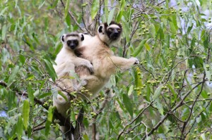
Verreaux Sifaka
Most of the island’s original forests have long been destroyed. With a growing population already over 22 million, and extensive rural poverty, the few isolated strips and pockets of forest that comprise the lemurs’ habitats are under increasing threat from slash-and-burn agriculture and from illegal logging of precious hardwood trees.
The current 2012-14 IUCN list of the world’s 25 most endangered primates includes six lemur species:
Blue-eyed Black Lemur (Eulemur flavifrons) – Endangered
Much needs to be done to strengthen the efforts of local organisations in Madagascar to raise awareness of the unique lemur plight and to help people find alternative means of making a living.
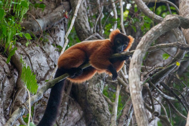
Red Ruffed Lemur
But Janet explains that there is also some good news. Conservation organisations like the Durrell Wildlife Conservation Trust are working with local people in Madagascar to help preserve Lemurs’ habitats, and there are some notable success stories.
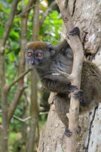
The ‘Bandro’, or Bamboo Lemur
In the Lake Alaotra area villagers have been helping to protect wetland reed-beds from destruction. The ‘Bandro’, or Bamboo Lemur (Hapalemur alaotrensis), which depends on the reed-beds for food and shelter, is no longer on the Critically Endangered list.
There are many fascinating displays in the Museum, but there’s something special about meeting an expert and chatting to them about the collections they love. Every Monday to Thursday our Spotlight Specimens series gives you the chance to do exactly that.
Taking place under the T. rex in the Main Court at 2.30pm each day, staff from across the collections choose favourite specimens to share with the public. These experts will also be writing a series of Spotlight Specimens blog posts for those of you who can’t make it to the Museum to meet them in the flesh. In this, the first in the series, Gina Allnatt kicks us off with a Halloween special…
**
It’s October, month of falling leaves and trick-or-treating, so what better way to get into an autumnal mood than to talk about two moths with marvellously morbid names?
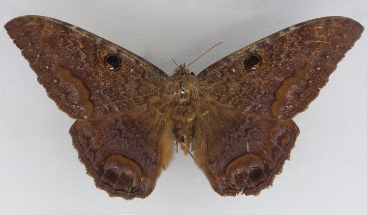
What do the Death’s Head Hawkmoth (Acherontia styx) and Black Witch moth (Ascalapha odorata) have in common? They are both associated with the film and novel Silence of the Lambs. The Death’s Head was used in the film, but the moth in the novel was originally the Black Witch. The moth was changed for the film for two reasons: The producers thought that a moth with a skull on its back would look more sinister, and also because it was almost impossible to get live specimens of the Black Witch moth for filming.
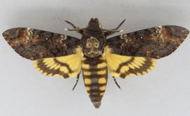
Death’s Head Hawkmoth (Acherontia styx)
The author of Silence of the Lambs, Thomas Harris, may have chosen the Black Witch moth because of the many legends and myths that surround it. In Jamaica it is known as the “Duppy Bat.” In Central and South America it’s known as “Mariposa de la Muerte”, which translates as “Butterfly of the Dead” because there is a myth which claims the moth is a harbinger of death. A less sinister version of this myth suggests that if you find one of these moths in your home it means an ancestor or loved one who recently passed away is paying you a visit.
However, the subtlety of these myths would probably not translate so well on film, so Mr. Death’s Head Hawkmoth took centre stage. The vernacular name of this moth comes from the skull-like markings on its back. There are actually three species of Death’s Head Hawkmoth- A. atropos, A. styx and A. lachesis. Though the moth mentioned in the film is Acherontia styx, Acherontia atropos was actually used instead.
All Acherontia supplement their diet by raiding the hives of bees for honey. The moths achieve this by using their extremely thick cuticle, which makes them impervious to stings. But the moth also uses another tactic: it is able to emit an odour that is chemically identical to the worker bees’ scent. This fools the bees into thinking the moth is one of their own. They also emit squeaking noises while in the hive. Some scientists posit that the squeak is similar to the noise a queen honeybee emits when she wants the workers to freeze. No one has been able to observe this theory, however.
Black Witch moth (Ascalapha odorata)
Despite all the myths and legends surrounding the Death’s Head Hawkmoth and Black Witch moth, both are large and harmless species. It is perhaps the fact that most moths are nocturnal which gives rise to so many legends and misinformation about them. It’s often the case that people will love butterflies but don’t like moths. Moths evolved before butterflies, and it is likely that the butterflies people hold dear evolved from day-flying moths (many day-flying moths exist today and are even more colourful than their butterfly counterparts!).
So remember this when you next see a moth (the original butterfly!) fluttering near a lamp as the sun slowly disappears.
Gina Allnatt, Curatorial assistant (Lepidoptera)
Gina will be talking about the Death’s Head Hawkmoth and Black Witch moth at 2.30pm on 28 and 31 October as part of our Spotlight Specimens series, running Monday to Thursday at 2.30pm.
The ecological importance of bumblebees has become more widely appreciated in recent years, thanks to environmental campaigners and reports of species decline, and even some extinctions, in the UK.
To look at this issue, we have recently teamed up again with arts-science organisation Pale Blue Dot, which is launching a new research project to investigate why some species of bumblebee are declining and to raise awareness about the ultimate impact this has on people.
Here, Pale Blue Dot co-founder Jane King explains how the Bees & Weeds project brings together art students, public engagement, the Museum’s collections and a leading bumblebee scientist…
*
On 9 September we launched our latest project – Bees & Weeds – with the Museum of Natural History, building on our previous collaboration for the Lost & Found exhibition. We were joined by over 50 art students from Banbury & Bicester College to highlight the plight of the bumblebee, revealing how its decline is impacting everything from what we eat to where we live and work.
Amoret Spooner displays drawers from the collections
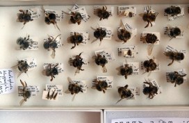
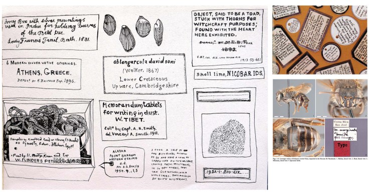
The students spent some time looking at methods of insect labelling and notation, before heading behind the scenes with entomologist Amoret Spooner to the Huxley Room, the location of the Great Debate on Darwin’s Theory of Evolution, which took place in 1860.
Amoret provided an insight into taxonomy – the science of species classification – as well as her work on the conservation of specimens. We visited the huge archive of bee specimens and learnt about some of the research that scientists are currently carrying out on UK bumblebee species to help prevent further decline.
Student sketches of labelling and notation
During spring 2015, the art students from the Banbury & Bicester College, as well as students from Oxford Brookes University, will make and install hundreds of cycle seat covers on bikes in and around the Oxford city. The seat covers will carry messages about bumblebee decline in the UK countryside, showing how much we depend on their pollination services, which far outstrip those of the honeybee in their value to UK food production.
We are also working with Professor Dave Goulson from the University of Sussex, one of the world’s most important bumblebee scientists. He will present his research showing how well bumblebees are doing in gardens compared to the countryside, as well as the optimum range of flowering plants needed to help them thrive. Dave’s book, A Sting in the Tale, is already a best-seller, and the sequel – A Buzz in the Meadow – was published on 4 September.
Dave will also be speaking about his new book at the Museum on Thursday 9 October at 7pm. Book your tickets for that via Waterstones here.
Artwork from the Bees & Weeds project, together with cycle seat covers and bike paraphernalia, will be on show and for sale in the Old Fire Station in Oxford from next spring. If you cycle in Oxford, you may be lucky enough to receive one!
Pale Blue Dot is an arts-science organisation helping scientists to communicate their research to the public. It promotes an interdisciplinary approach to learning, living and working through exhibitions, publications and happenings.
They say a picture is worth a thousand words, and at the Museum we make thousands of pictures: pictures to document, pictures to investigate, and pictures to wow. We use a lot of different imaging techniques too, from standard close-up photography to scanning electron microscopy, which reveals the most minute details.
To coincide with the final week of the Wildlife Photographer of the Year exhibition here, on Saturday 20 September we held a new adult workshop to give people some hands-on experience of some of these processes. Imaging Techniques in Modern Natural History gave participants the chance to get up close to some wonderful specimens and make their own images to take home.
I had planned to review the day here, but Rose Parkin, who took part in the workshops, very helpfully sent in her own write-up of the sessions. So here’s a special guest post from Rose, along with some pictures taken by people on the day.
*
By Rose Parkin
When I signed up for the digital imaging course I expected a fairly dry, tech-heavy day. Instead, the experience was really exciting. Not only did it provide hands-on experience of viewing and recording images with new technology, it also gave me a brief glimpse behind the scenes of my favourite museum.
Laser Scanning and Digital Modelling For our first session our small group was led through a maze of corridors by Sarah Joomun, the Documentation Officer, to the laser scanning lab. It sounded a bit futuristic, and it turned out that it looks that way too. Sarah popped a fossil onto a mount, clicked a few buttons and red lasers appeared, scanning the fossil’s surface while it rotated. After ten minutes the first 3D image of the fossil was produced – a beautiful net of triangles, which looked like a teleporting object in a science fiction film.
Laser scanning Image copyright Tom Nicholson-Lailey
Sarah turned the fossil and scanned it again. The challenge was then to fit these two images together to make a complete 3D model. Amazingly, this technique enables other palaeontologists around the world to see and replicate, with the use of a 3D printer, the exact size and shape of a fossil without it ever leaving the museum.
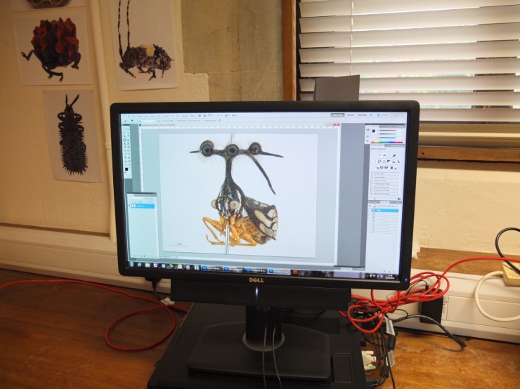
Multi-plane Microscope Photography Our next session was upstairs, with artist-in-residence and photographer Katherine Child. Even though we were close to the main corridor of the museum it felt like a real working space, crammed full of equipment and insect specimens. Katherine had chosen the tiniest of insects for us to photograph with the multi-plane microscope. It looked like a small seed with some barely visible limb-like protrusions.
Multiplane photography. Image copyright Rose Parkin
But under the microscope a wonderfully strange insect became visible, with the most bizarre appendages and bright orange legs. While the microscope already showed a great deal of detail the multi-plane photography captured an incredibly crisp image. The microscope takes large numbers of photos of the specimen, using different focal planes each time, then the focussed elements are all stacked together to produce a crystal clear photograph.
Once we’d chosen and photographed some other insects from the collection and poked around the room a bit (finding a disturbing collection of large pickled spiders), we were taken on a tour of the entomology department. Katherine led us through corridors of offices and labs, up to a stunning store room that felt almost church-like, with rows and rows of cabinets full of fascinating insects.
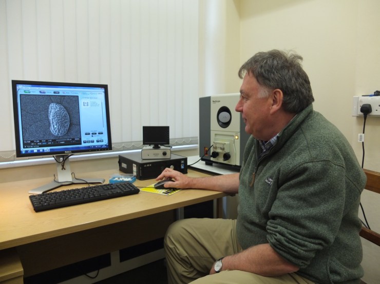
Scanning Electron Microscopy After lunch we had a laboratory session with museum director Paul Smith to look at sand under an electron microscope. Luckily, that was much more exciting than it sounds! The sand was taken from Dog’s Bay on the west coast of Ireland and is rich with a wide range of tiny fossilized organisms. Paul showed us how to carefully select individual microfossils from a tray using just a microscope and a paint brush.
Professor Paul Smith demonstrates the scanning electron microscope. Image copyright Rose Parkin
We then viewed some of the microfossils using a scanning electron microscope. This allowed us to see an incredible level of detail. The microscope was so powerful that we could see hair holes in a fossil the size of a grain of sand.
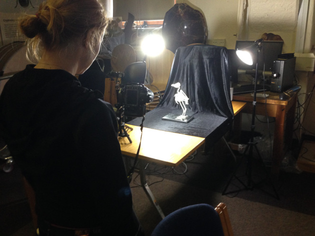
DSLR Macrophotography My final session was a crash course in macrophotography. Held in the seminar room, the low lighting and floor-to-ceiling collection of specimens lent an almost eerie feeling to the set-up.
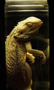
Macrophotography. Image copyright Keith BarnesBearded dragon. Image copyright: Rose Parkin
Once prepped, we were let loose on four separate camera setups. Being able to choose and shoot at our own pace made this feel like a really creative experience. The help given by professional photographer Keith Barnes and public engagement officer Scott Billings was perfect – very hands on but not patronizing (despite my lack of DSLR experience).
With this digital imaging course the museum has created a really exciting snapshot of the work that goes on behind the scenes, reinforcing the fact that this impressive place is much more than just an ordinary museum.
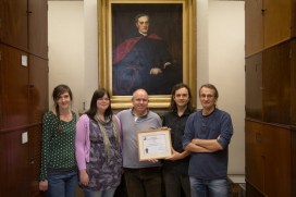
Katherine Child, Kate Santry, Peter Eeles, James Hogan and Darren Mann (l-r)
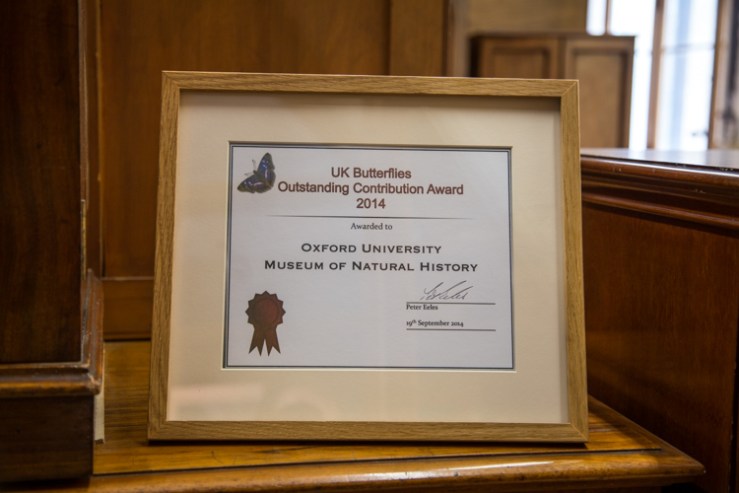
Congratulations to our fabulous Life and Archival Collections teams! They’re a pretty friendly bunch, always keen to help researchers and enthusiasts, giving unrivalled access to their knowledge and their collections. This welcoming attitude has now been recognised by the organisation UK Butterflies, who have awarded them with an Outstanding Contribution Award.
Peter Eeles (l) presents the award to James Hogan
This is the first time that the award has been given to an organisation, rather than an individual, which was apparently “in special recognition of the role that this team of committed individuals has played in helping bring the UK Butterflies website to a whole new level.”
Peter Eeles, who developed and runs the UK Butterflies website, has been visiting the Hope Entomological Collections (part of our Life Collections) for many years. He says that the staff have always been “encouraging, knowledgeable and welcoming” and have truly helped advance the mission statement of the organisation; Building a community of responsible butterfly enthusiasts.
Peter admires some Camberwell Beauty butterflies.
James Hogan, from the Hope Collection, has worked closely with Peter and his colleagues and says;
“It’s a great honour to be recognised and it’s always a pleasure to welcome the UK Butterflies members.
“They’re always enthusiastic about what they do and I learn a huge amount about our collections, too. There’s no point having collections if no-one uses them!”
The Library and Archives, run by Kate Santry, has been vital in research into the history of entomology. Last year, Mark Colvin, a contributor to the website, used the collections to study the life and works of James Charles Dale. By combining diaries and photos from the Archives and specimens from the Life Collections, he built up a thorough report on the entomologists’ work. Peter interviewed Kate about her work back in December last year and put the interview onto the website.
Mark and Peter talked about the impressive ‘open door’ policy that the staff have, and this doesn’t just extend to entomologists. Kate is always very keen to point out that the Library is open to anyone who would like to visit. Just drop her an email on libary@oum.ox.ac.uk to arrange a good time to call in.
Thanks to Peter, Mark and all at UK Butterflies, for this exciting award – now to find the perfect spot to show it off!
Rachel Parle, Interpretation and Education Officer






 There are many fascinating displays in the Museum, but there’s something special about meeting an expert and chatting to them about the collections they love. Every Monday to Thursday our Spotlight Specimens series gives you the chance to do exactly that.